Cellular BRD4 Protein Degradation in PROTAC dBET6 Treated HepG2 Cells by Western Blot
BRD4
BRD4
BRD4
HepG2 cells from ATCC
Western Blot
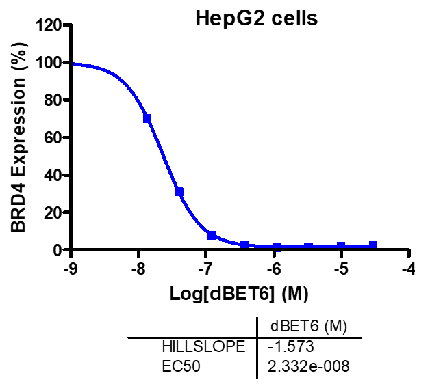
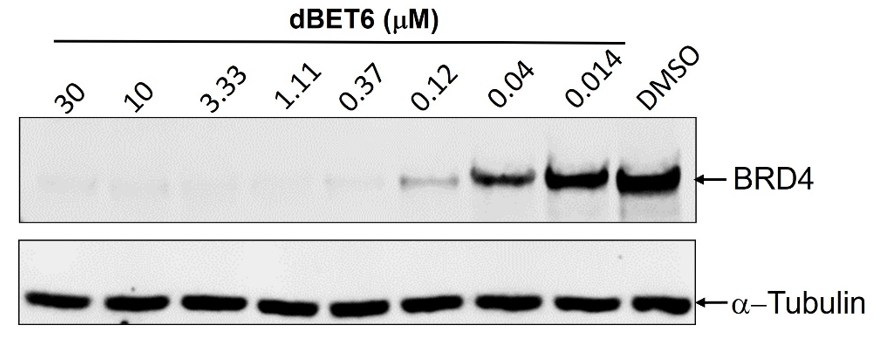
HepG2 hepatocellular cancer cells were seeded into the wells of 12-well plates. The cells were treated with reference PROTAC compound dBET6 for 8 hours. The cell lysates were subjected to Western blot analysis. The blots were probed with BRD4 antibody and α-Tubulin antibody. The membrane was scanned with LI-COR Odyssey Fc Imaging System. The specific bands of interest were quantified by LI-COR Image Studio Lite software. The IC50 curve was plotted and IC50 value was calculated using the GraphPad Prism program based on a sigmoidal dose-response equation.
LI-COR Odyssey Fc Imaging System
| Compounds | IC50 (M) |
|---|---|
| dBET6 | 2.332e-008 |
Malvern, PA, USA
More information can be found on our website Protein Degradation Assay – PROTAC Screening
Western blot for detection of BRD4 degradation in PROTAC dBET6 treated HepG2 cells
Western blot quantification for IC50 determination of PROTAC dBET6 induced BRD4 degradation