FLT3 (wild type control) Cellular Phosphorylation Assay (intracellular kinase activity assay) for compound screening and profiling in intact cells
The receptor tyrosine kinase FLT3-wt (FMS-like tyrosine kinase 3 (also known as Flk2 for Fetal liver kinase 2) belongs to the class III receptor family. Expression of FLT3-wt is found in early myeloid and lymphoid precursor cells and plays a crucial role in the development of the haematopoietic system. In 70-90% of all AML/ALL patients leukemia cells display increased FLT3-wt levels. In some of these cases activating internal tandem duplications (ITDs) or point mutations within the FLT3 gene were identified. The most common activating point mutation is an exchange of aspartate 835 to tyrosine (D835Y) which is found with a frequency of 60% in AML patients with point mutated FLT3.
FLT3
FLK2, STK1, CD135
MEF
Transfected
Reaction Biology’s cellular FLT3-wt phosphorylation assay was generated on a mouse embryonal fibroblast (MEF) background. Cells were transfected to express a full-length FLT3-wt molecule. Binding of FLT-3 ligand (FLT3-L) activates receptor autophosphorylation via the mechanism of receptor dimerization. After stimulation with FLT3-L phospho-FLT3 levels are determined by a sandwich ELISA system. The assay is validated based on known inhibitors of FLT3-wt kinase activity (see Fig. 1).
Substrate phosphorylation as a readout of intracellular kinase activity via ELISA
Freiburg, Germany
More information can be found on our website Cellular Phosphorylation Assay Services.
Primary reference compound IC50 for FLT3 (wild type control)
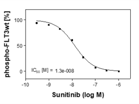
Sunitinib[1] is a potent inhibitor of the FLT3-L induced phospho-FLT3-wt signal found in the described cells. The graph shows a representative result.
[1] O’Farrell, AM et al. (2003) Blood 101, 3597-3605
Additional validation data

Staurosporine[1] is a potent inhibitor of the FLT3-L induced phospho-FLT3-wt signal found in the described cells. The graph shows a representative result.
[1] O’Farrell, AM et al. (2003) Blood 101, 3597-3605