NK Cell Killing Assays
Immuno-Oncology Platform

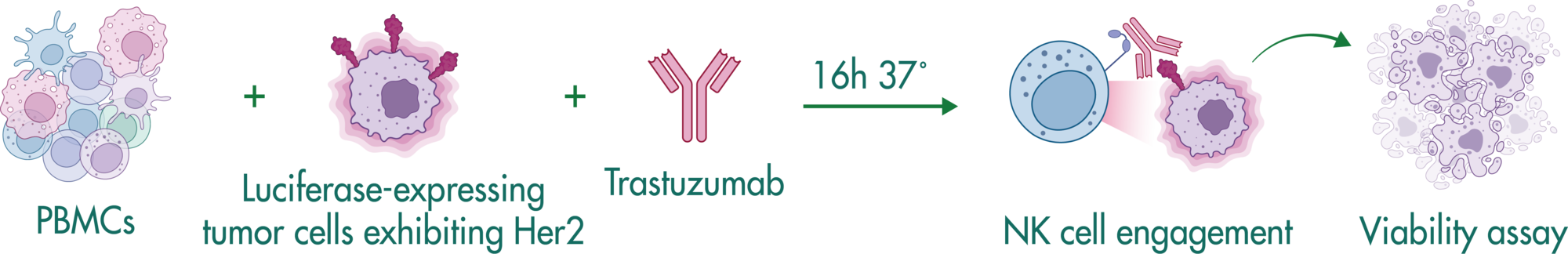
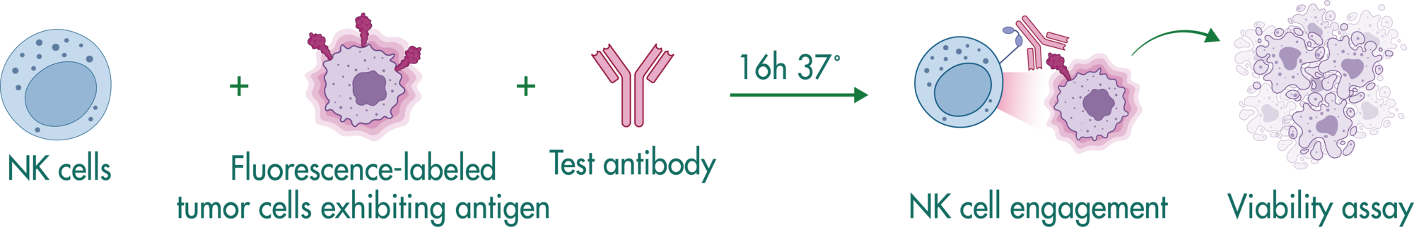
NK Cell Killing Assays (ADCC Assay)

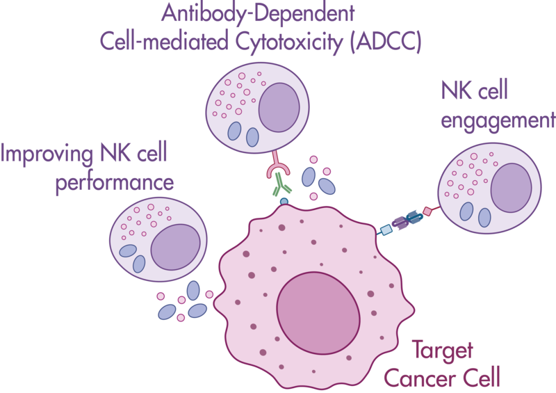
NK cells can recognize and kill cells that show signs of oncogenic transformation. This unique functionality puts NK cells in the center of immunotherapy research seeking to help NK cells identify tumor cells and enhance the therapeutic activity of NK cells.

Natural killer cells are regulated by activating and inhibiting receptors. Tumor cells express stress-induced ligands or lack sufficient MHC-I molecules, which are recognized by NK cells prompting the release of cytotoxic granules. The innate immune process antibody-induced cellular cytotoxicity (ADCC) also involves NK cells. They express Fc receptors on their surface, recognizing and killing antibody-coated target cells. Many advantages of NK cell therapies are based on their innate nature; for example, cells can be transferred between individuals, opening opportunities for off-the-shelf products.

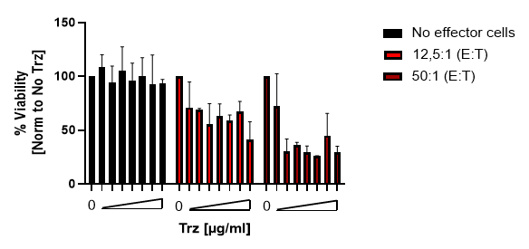
Reaction Biology offers a variety of immune cell assays. Our ADCC assays enable the investigation of antibody treatment sought to recognize malignant cells and label them for killing by NK cells. Get in contact with us to have a conversation with our scientists about your research goals and how we can help you achieve them.

NK Cell Killing Strategies for Immunotherapy

Several strategies aim to improve tumor reduction via NK cell killing:

  • Enhancing NK cell killing performance by augmenting NK cell function
  • Antibody-dependent cellular cytotoxicity (ADCC)
  • NK cell engagement via bi-specific molecules
  • Increasing the number of NK cells in patients by adoptive transfer

NK Cell Killing Assays Available at Reaction Biology

  • Assay options
  • Study example: ADCC assay performed with PBMCs
  • Study example: ADCC assay performed with isolated NK cells
  • Study example: NK engager molecule testing
Assay options

Reaction Biology offers a versatile suite of immuno-oncology assay setups. Since every project is different, we enable a customized study layout with various options for immune cells, target cells, or readouts.

Please inquire about further customization requirements to tailor an assay specific to your research needs.

Options for immune cells

  • PBMCs
  • isolated NK cells

Options for target cells

  • human or murine tumor cells
  • tumor cells stably expressing tumor-associated antigens or luciferase
  • 2D or 3D cell culture

Options for readout

  • quantification of luciferase-expressing tumor cells
  • flow cytometry
  • live-cell imaging and high-content imaging
  • Bliss factor determination of drug combination therapies

Readout options

  • NK-mediated ADCC assay
  • NK cell killing mediated by NK cell engager molecules
  • further readout options are available upon request
Study example: ADCC assay performed with PBMCs

Approach

In this study example, we explore the effect of the monoclonal anti-Her2 antibody Trastuzumab (Trz) on NK cell killing by co-cultivating PBMCs with tumor cells expressing the cognate antigen.

Setup


Luciferase-expressing tumor cells exhibiting Her2 are seeded in a 384 well plate. PBMCs at different E:T (effector : target cell) ratios are added on top of the tumor cells. After an incubation period of 16 hours in the presence of Trastuzumab, the luciferase activity is measured as a readout of tumor cell viability.

Study results

Trastuzumab (Trz)-induced immune killing of luciferase-labeled SKOV-3 tumor cells.


PBMCs from a healthy donor were incubated at different E:T ratios in the presence of various Trz concentrations with the Her2-expressing cell line SKOV-3-FireLuc. After 16-18h incubation, luciferase activity was measured, and the % viability was calculated using 1% Triton-treated cells as maximal cell death and untreated SKOV-3-FireLuc as maximal viability. The bar graph shows the mean and SD of % viability normalized to the untreated (no Trz) condition.

Study example: ADCC assay performed with isolated NK cells

Approach

In this study example, we explore the effect of a monoclonal antibody on NK cell killing by co-cultivating NK cells with tumor cells expressing the cognate antigen. Viability testing of the tumor cells was performed via flow cytometry.

Setup

NK cells were isolated from healthy donor PBMCs with a human NK cell isolation kit. Target cells were co-cultivated at different ratios for 4 hours in the presence of a monoclonal antibody recognizing a target cell-specific antigen. Tumor cells were detected via CellTracker Green dye and viable cells were quantified using a viability dye by flow cytometry.

Study results

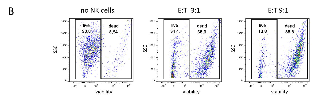
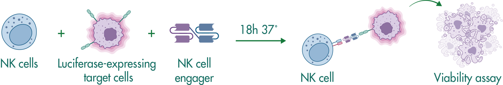
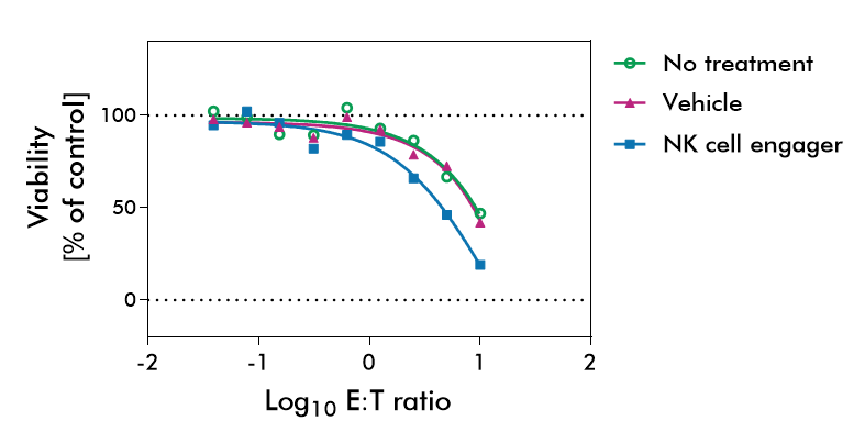
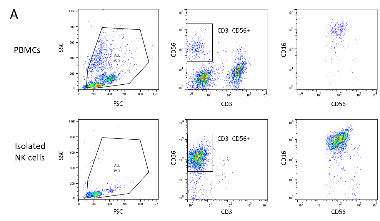
NK cells were isolated from healthy donor PBMCs via an NK Cell Isolation Kit. The purity of isolated NK cells was quantified by staining with NK cell-specific markers CD3-, CD56+, CD16+  before isolation (upper row) and after isolation (lower row). 10 million NK cells could be recovered from 300 million PBMCs with 88% purity.

Viable target cells were quantified after a 4-hour co-cultivation of isolated NK cells and target cells in the presence of a target cell-specific antibody. The target cells were stained with CellTracker Green and subjected to viability testing with a live/dead dye via flow cytometry.

The presence of NK cells in a ratio of 3:1 increased the number of dead target cells from 9% to 65%. A ratio of 9:1 resulted in 86% of dead target cells.

Study example: NK engager molecule testing

Assay principle

Natural killer cell engagers are designed to tether NK cells to tumor cells. These multi-specific antibodies contain fragments specifically binding to NK cells and to tumor-associated antigens facilitating tumor cell killing.

Setup

NK cells were isolated from healthy donor PBMCs with a human NK cell isolation kit. The luciferase-expressing tumor target cells were co-cultivated with the NK cells at different ratios overnight in the presence of a bi-specific antibody recognizing NK cells and a tumor cell-specific antigen. At the end of the incubation period, the cells were lysed, and luciferase was quantified as a readout of tumor cell viability.

Study results

Co-cultivating NK cells and tumor target cells resulted in a decline of tumor cell viability in the presence and absence of the NK cell engaging test substance due to the innate function of NK cells to recognize and kill tumor cells. In the presence of a test substance, however, the tumor cell killing could be augmented significantly. At an effector:target cell ratio of 10:1, only 35% of tumor cells were viable in the presence of the NK cell engager compared to around 50% viable tumor cells in the control groups.