Apoptosis Assays
Cell-Based Assays

Apoptosis Assay Services for Drug Discovery

Apoptosis assays elucidate the cell death-related events during tumor cell killing by anti-cancer drugs. Two key questions need to be answered: Do tumor cells die? And, through which pathway do the cells die?

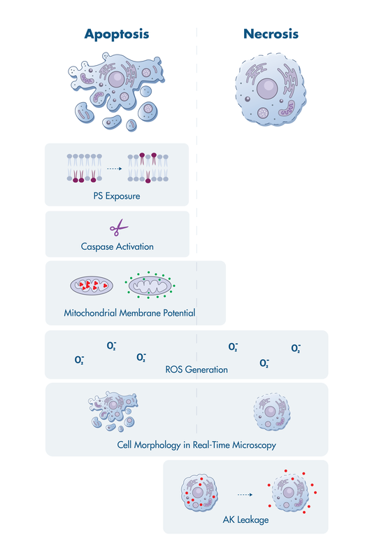
Reaction Biology offers a suite of cell death assay services to investigate events that have been linked to apoptosis including activation of mitochondrial membrane permeabilization, caspase activation, surface exposure of phosphatidylserine, and more.

Apoptosis assays are available for single and multi-time point analysis as well as real-time live-cell imaging.

Cell Death Assays

Apoptosis is the regulated process of cellular dismantling that avoids eliciting inflammation. Necrosis is a passive cell death with perforation of the plasma membrane and uncontrolled release of inflammatory cellular contents.

Both forms of cell death have distinct characteristics that can be monitored in a variety of assay formats at Reaction Biology.

PS … phosphatidylserine
ROS … reactive oxygen species
AK… adenylate kinase
Image adapted from O. Kepp et al., NRDD, 2011.

cell death mechanisms

Apoptosis Assay Details

  • Caspase 3/7 Activity
  • Caspase 3 Cleavage
  • Annexin V Staining
  • Mitochondrial Membrane Potential
  • ROS Quantification
  • AK Leakage
Caspase 3/7 Activity

Principle: The activation of the caspase cleaving cascade is one hallmark of the apoptosis pathway. Caspase 3 and 7 are effector caspases executing apoptosis events by cleaving a large set of substrates resulting in the regulated destruction of cells.

Assay format: For detection of caspase 3 and caspase 7 activity, we use Promega’s Caspase-Glo 3/7 Assay system, which is a homogenous assay with a simple add-mix-measure protocol enabling testing in high-throughput capable 384 well format.

The Caspase-Glo 3/7 substrate is cleaved by both caspase 3 and caspase 7, resulting in free aminoluciferin, which is consumed by luciferase resulting in a luminescent signal for quantification by an Envision Multilabel Reader.

Real-Time Imaging:  Our IncuCyte Live-cell Analysis system enables real-time, automated measurement of caspase 3/7 activity. Sartorius’ IncuCyte Caspase 3/7 Dyes comprise caspase substrates to release a DNA-binding fluorescent label.

View Caspase 3/7 Activity example study

Caspase 3 Cleavage

Principle: The detection of the activation of caspase 3 by proteolytic processing.

Assay format: Tumor cells are lysed, and the lysate is subjected to SDS-PAGE to separate the proteins in the lysate by their molecular size. Cleaved and uncleaved caspase 3 are detected via western blotting by an anti-caspase 3 antibody. The proteins are quantified with a LICOR Odyssey Infrared Imager.

View Caspase 3 Activation example study

Annexin V Staining

Principle: During apoptosis, the loss of plasma membrane symmetry results in the appearance of phosphatidylserine on the outer plasma membrane, which serves as a signal for the uptake of apoptotic bodies by macrophages.

Assay format: Annexin V staining is a simple homogenous add-mix-measure assay. Briefly, cells incubate in multiwell plates and are treated with the test compound. After incubation, the cells are detached from the well surface by accutase treatment. Fluorescence-labeled Annexin V is added to the cell culture, and apoptotic cells are quantified with the Envision Multilabel Reader.

For additional information, the membrane-impermeable propidium iodide can be added to distinguish viable apoptotic cells from necrotic cells.

Mitochondrial Membrane Potential

Principle: During apoptosis, the electrochemical gradient across the mitochondrial membrane, a prerequisite for ATP production, collapses due to the mitochondrial permeability transition event. The visualization of the integrity of the membrane potential is enabled by the cationic dye JC-10, which accumulate in healthy mitochondrial membranes in proportion to the membrane gradient ΔΨm to form red fluorescent aggregates. In apoptotic or necrotic cells, the dye diffuses out of the mitochondria forming monomers emitting green fluorescence.

Assay format: The assay works with a simple mix-add-read format. The JC-10 dye is added to the tumor cells. After the designated incubation time, the cell culture plate is read in the Envision Multilabel Reader quantifying red and green fluorescence.

Real-Time Imaging: Our IncuCyte Live-Cell Analysis setup allows monitoring of the mitochondrial membrane potential in real-time. Please inquire for more information.

View Mitochondrial Membrane Potential example study

ROS Quantification

Principle: The accumulation of reactive oxygen species (ROS) such as H2O2 is one facilitator of cell damage and cell death detected in both apoptosis and necrosis.

Assay format: ROS detection is performed with the ROS-Glo H2O2 Assay from Promega. This simply add-mix-read assay contains a luciferin derivate as substrate, directly reacting with H2O2 to generate a luciferin precursor. This precursor is converted to luciferin, which can be quantified in a luminescent luciferase reaction with the Envision Multilabel Plate reader.

AK Leakage

Principle: Adenylate kinase (AK) is an intracellular enzyme present in all cells. Upon loss of cell integrity by disruption of the plasma membrane, for example during necrosis, AK is released in the cell medium. Quantification of AK release allows accurate determination of cell lysis and cytotoxicity.

Assay format: To detect AK in the cell medium we use Lonza’s ToxiLight Cytotoxicity Assay kit. The measurement is based on two steps, first, ADP is added to the cell medium as a substrate of the enzyme AK generating ATP. Second, the newly generated ATP is measured via a bioluminescent luciferase reaction.

Advantage: Because the assay needs only small amounts of cell medium, a disruption or lysis of the cells is not necessary. This enables investigation of cytotoxicity kinetics and other readouts, for example, of markers of apoptosis or cell cycle.