T Cell Killing Assays
Immuno-Oncology Platform

T Cell Killing Assays

T cell killing assays measure a drug’s potency to help T cells kill tumor cells. T cell killing assays are a valuable tool for immuno-oncology discovery projects for in vitro assessment of new drug candidates to:

  • Understand the immune-modulating action of new immunotherapeutics
  • Optimize the potency of new drugs
  • Select the most promising drug candidates for animal studies.

Cytotoxic T cells can recognize and kill target cancer cells. New therapies are designed to facilitate T cell activation and increase effector function while suppressing inhibitory mechanisms. Another approach is helping T cells to move in the proximity of their target cells. Both approaches can be investigated with our T cell killing assays. We are also open to exploring further avenues of modulating the immune response with our assays.

Our T cell killing assays can be used in single substance or drug combination modality.

T Cell Killing Strategies of Immunotherapies

Various strategies are used to enable tumor cell killing by cytotoxic T cells:

  • Blockage of immune checkpoint inhibition
  • T cell activation via binding to anti-CD3 and other stimulating receptors
  • Physically engaging T cells and tumor cells
  • Creation of T cells expressing tumor cell-specific T cell receptors (CAR-T)

Reaction Biology’s killing assays were created to test the efficacy of these immunotherapy strategies on T cells.

T Cell Killing Assays Available at Reaction Biology

  • Assay options
  • ADCC assay performed with Jurkat T cells stably expressing FcγRIII receptor and an NFAT response element
  • Study example: Complement Dependent Cell Cytotoxicity (CDC) assay
  • Study example: Anti-CD3+anti-PD1 treatment
  • Study example: BiTE induced killing of lymphoma cells
Assay options

Reaction Biology offers a versatile suite of immuno-oncology assay setups. Since every project is different, we enable a customized study layout with various options for immune cells, target cells, or readouts for our customers to choose from.

Please inquire about further customization requirements to tailor an assay specific to your research needs.

Options for immune cells

  • PBMCs
  • isolated T cells

Options for target cells

  • human or murine tumor cells
  • tumor cells stably expressing tumor-associated antigens or luciferase
  • 2D or 3D cell culture

Options for readout

  • quantification of luciferase-expressing tumor cells
  • flow cytometry
  • live-cell imaging and high-content imaging
  • Bliss factor determination of drug combination therapies

Therapy options

  • T cell killing enhanced by T cell activation
  • T cell killing mediated by T cell engagement
  • further therapy options are available upon request
ADCC assay performed with Jurkat T cells stably expressing FcγRIII receptor and an NFAT response element

Assay principle

Components of the cell-mediated immune system target and kill virus-infected or other diseased (e.g., cancer) cells. When FcR-bearing effector cells identify a target cell that has been opsonized by antibodies, the ADCC pathway is activated. Once the effector cells have been activated by binding, the target cells will be killed by the cell-mediated ADCC immune defense mechanism.

Goal

Antibody-dependent cell-mediated target killing of virus-infected or other diseased cells.

Setup

Effector cells (Jurkat T cells) cultured for ADCC bioassays are engineered with stably expressing FcγRIIIa and have an NFAT response element that promotes the expression of firefly luciferase. Upon antibody binding to target antigens on the cell surface of the target cells (tumor cells, virus-infected cells, or other diseased cells), the Fc portion of the target-bound antibody binds to the FcγRIIIa receptors on the cell surface of the effector cell. Through multiple cross-links, the binding ensures precise cell-to-cell recognition, which ultimately results in ADCC activation. The result of activating this pathway leads to the death of the target cells.

Example Data

ADCC Reporter Bioassay was used to measure the Fc effector function of Rituximab to demonstrate assay specificity.

Study example: Complement Dependent Cell Cytotoxicity (CDC) assay

Introduction

TNF is involved in the pathology of several inflammatory and autoimmune diseases, and the use of neutralizing antibodies or engineered TNFR fusion proteins to block TNF/TNFR binding has proven to be a viable strategy for providing clinical benefits in inflammatory diseases. TNF-producing cells could be targeted for destruction, which could help reduce the inflammatory burden even more. TNF-targeted antibodies can activate effector functions such as antibody-dependent cell-mediated cytotoxicity (ADCC) or complement-dependent cytotoxicity (CDC) to destroy mTNF-expressing inflammatory cells.

Goal

Complement-dependent cytotoxicity (CDC) assays are conducted to evaluate new therapeutics during the drug development phase. The CDC assay is used for safety profiling by screening therapeutic candidates to ensure that the compound does not induce complement events and to look for other unexpected effects. Fresh blood that is less than four hours old is used in this assay, as it generates better responses and results.

Example Data

CDC activity of infliximab assessed using the CDC assay developed by Reaction Biology.

Study example: Anti-CD3+anti-PD1 treatment

Killing of luciferase-expressing tumor cells via PBMCs stimulated with anti-CD3 treatment enhanced by immune-checkpoint inhibition

Assay principle

Engaging the T-cell receptor complex leads to activation of resting T cells and induction of cytokine release. In this study example, we determined the potency of drugs enhancing T cell activation by co-cultivating PBMCs with tumor cells in the presence of anti-CD3. Further, we modulated the T cell’s killing capacity with the immune checkpoint inhibitor anti-PD1.

Setup

Tumor cells stably expressing luciferase were co-cultured with PBMCs at an E:T (effector to target cell ratio) of 6:1 or a range of E:T ratios, in the presence of anti-CD3 at low concentrations to induce killing at a suboptimal level. In a second experiment, the checkpoint inhibitor anti-PD1 was added at different concentrations (7x semi-log). The co-culture was incubated for 4 days after which the viability of the tumor cells was measured by luciferase activity.

Study results

T cell stimulation via anti-CD3 antibody resulted in activation of T cells and killing of tumor cells (A). This effect was enhanced by drug combination testing with anti-PD1 antibodies (B).

PBMCs from a healthy donor were co-cultured with cancer cell lines HT29 and COLO205 stably transduced with Firefly luciferase (FLuc).

A. An E:T ratio of 6:1 was used with the addition of semi-log dilutions of anti-human CD3 (OKT3).
B. Anti-human CD3 (OKT3) at a concentration of 0,68 ng/ml was used to stimulate T cells in a killing assay with different E:T ratios. Anti-PD1 antibody Nivolumab treatment slightly enhanced tumor cell killing.

The % viability was calculated using tumor cells alone as maximal viability (100%) and Staurosporine treatment as maximal killing (0%).

Study example: BiTE induced killing of lymphoma cells

Combination therapy to enhance BiTE-induced T cell killing of luciferase-labeled B-cell lymphoma cell line OCI-LY1

Assay principle

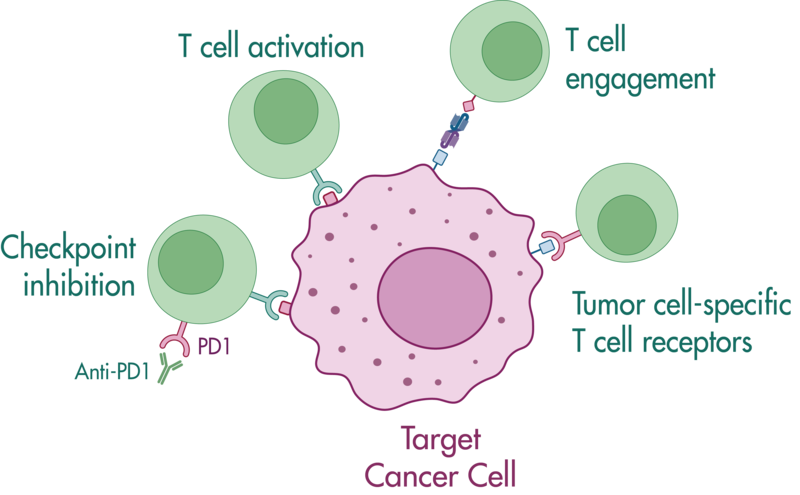
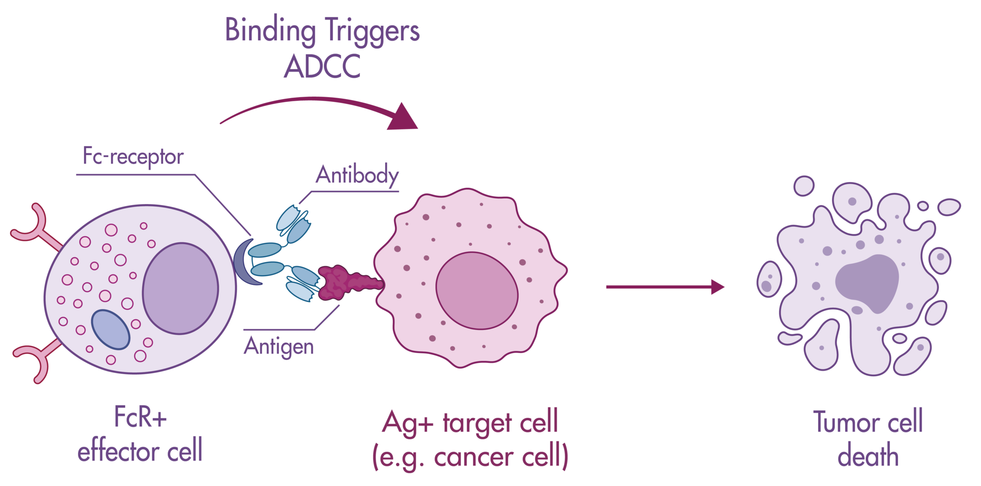
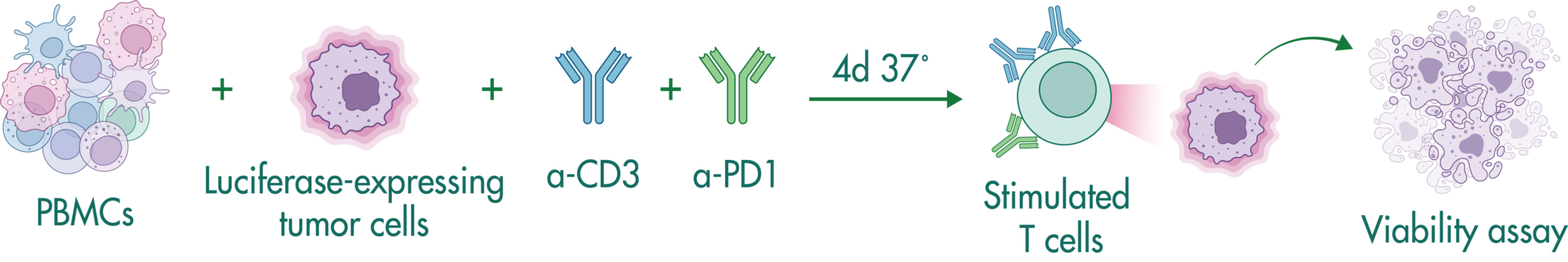
Bi-specific antibodies contain two binding sites to engage T cells and tumor cells resulting in tumor cell killing by (i) bringing the cells in close proximity, and (ii) stimulating cytokine release in T cells.

In this study example, we determine the capacity of a bi-specific engaging molecule to facilitate tumor cell killing by T cells and the combinatorial potential with other clinically relevant agents.

Setup

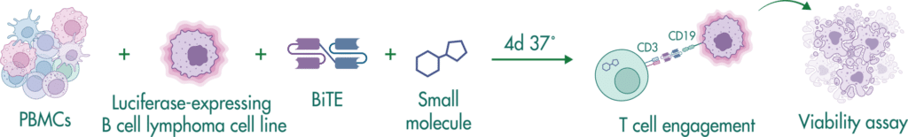
CD19-expressing B cell lymphoma cells stably expressing firefly luciferase are co-cultured with PBMCs at an E:T (effector to target cell ratio) of 2:1. We tested the bi-specific T cell engager Blinatumomab recognizing CD19 and CD3 at different concentrations (7x semi-log), with or without additional compounds. The co-culture was incubated for 4 days, after which the viability of the tumor cells was measured by luciferase activity.

Study results

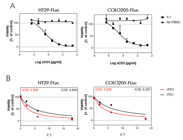
BiTE Blinatumomab and the small molecule Lenalidomide act synergistically to kill CD19-positive B cell lymphoma cells, whereas the combination with S63845 acts antagonistically.

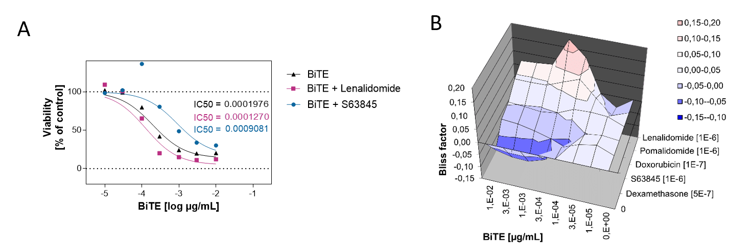
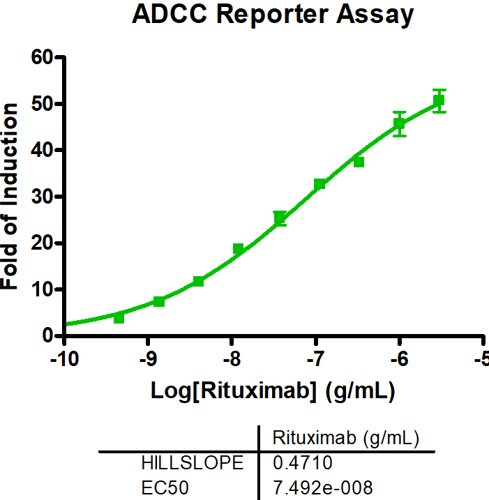
A. PBMCs from a healthy donor were incubated with the B cell lymphoma cell line OCI-LY1 at a 2:1 E:T ratio and treated with a range of 7 concentrations of Blinatumomab. Luciferase expression of live tumor cells was measured after 4 days, and viability was calculated using Staurosporine treated cells as maximal cell death control and untreated target cells as maximal viability control. An IC50 of 0,19 ng/ml was found for Blinatumomab. The combinatorial effects of Blinatumomab with Lenalidomide and MCL1-inhibitor S63845 are shown as examples.

B. Using the bliss factor scoring, we determined which drug combinations produce additive, synergistic, or antagonistic effects for killing tumor cells.