GPCR Assays
Target-specific Assays

GPCR Assays

G protein-coupled receptors (GPCR) represent the largest individual family of targets for currently approved medications. Recent advances in GPCR pharmacology, including biased signaling and allosteric modulators, have become increasingly important tools for GPCR assay development. Reaction Biology offers GPCR screening services to progress drug discovery research in the area of GPCR biology and pharmacology.

  • We offer GPCR assay development, high-throughput screening, and SAR support services.
  • Transmembrane signaling assay formats can be readily established for your receptor of interest including calcium mobilization, β-arrestin translocation, cAMP generation, and inositol 1-monophosphate (IP1) generation

GPCR signaling events used for drug screening

GPCRs are a large family of 7 transmembrane receptors transducing extracellular signals into intracellular responses. After agonist binding, GPCRs modulate the activity of downstream signaling enzymes such as adenylate cyclase which converts ATP to cAMP, a second messenger. Another well known GPCR pathway activates phospholipase C resulting in the generation of two second messengers: diacylglycerol (DAG) and inositol 1,4,5-trisphosphate (IP3) which is involved in intracellular calcium mobilization. A stable metabolite of IP3 is IP1 (inositol 1-monophosphate) which can be quantified. The translocation of β-arrestin is involved in the termination of GPCR signaling as well as modulation of additional signal transduction processes.

GPCR signaling

GPCR Assay formats

  • Custom-tailored screening platform
  • IP1 generation
  • cAMP generation
  • FLIPR Calcium Mobilization Assay
  • β-arrestin translocation
  • LinkLight™: Protein-Protein Interaction
Custom-tailored screening platform

Our dedicated team of GPCR experts will enable drug screening with assays tailored to your specific needs:

1. Define the needs and scope for the project together with our assay development team.
2. Make us familiar with the goals for your research project and define timelines to ensure goal-oriented work right from the start.
3. We will acquire or generate a cell line appropriate to the project needs.
4. The same high standards we use for our off-the-shelf assays will apply to newly developed assays for your project.
5. We guarantee fast turn-around times for data generation.
6. During every step of the process, you will be in close contact with your project manager for regular updates on the study progress.

IP1 generation

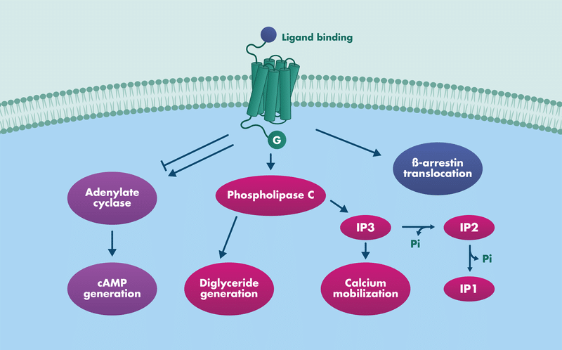
Cells engineered to express Gq-coupled receptors of interest are stimulated with a receptor agonist to elicit the generation of IP3. Given that IP3 is not metabolically stable, a surrogate marker of pathway activation (the metabolically stable IP1 (inositol 1-monophosphate) is measured. IP1 can be conveniently quantified using a TR-FRET-based competitive immunoassay as depicted above.


IP1 generation was quantified upon GPCR induction on an undisclosed agonist. Shown is also the IP1 standard curve for comparison.

cAMP generation

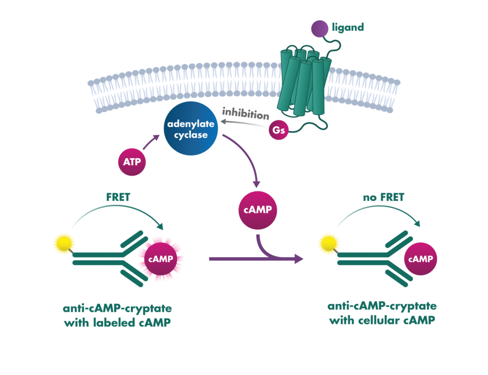
Cells engineered to express Gs-coupled GPCRs of interest are stimulated with an agonist to affect cellular activation. cAMP can be accurately measured by a variety of standard detection methods including a competitive immunoassay wherein cellular cAMP competes with a labeled cAMP probe to bind to an anti-cAMP-cryptate generating a FRET signal.


Levels of cAMP were measured after stimulation of a GPCR expressing cell line with an undisclosed agonist. A cAMP standard curve is used for data normalization.

FLIPR Calcium Mobilization Assay


The concentration-response of agonist stimulation for calcium mobilization is shown.

β-arrestin translocation

β-arrestins bind to activated GPCRs to mediate desensitization and internalization of GPCRs. They are scaffolding proteins that further mediate cell signaling pathways independent of G-proteins.

Example of β-arrestin translocation in response to treatment with an undisclosed agonist.

LinkLight™: Protein-Protein Interaction

LinkLight™: Protein-Protein Interaction


Cells engineered to express GPCRs of interest are stimulated with test compounds to trigger receptor activation. Upon ligand binding, the GPCR undergoes conformational changes leading to G-protein signaling followed by GRK-mediated phosphorylation. The phosphorylated receptor recruits β-arrestin, bringing TEV protease (fused to the GPCR) into proximity with permuted luciferase (fused to β-arrestin). TEV cleavage activates the luciferase, generating a persistent luminescent signal that can be accurately measured by standard luminometry detection methods.

Example curve

Levels of β-arrestin recruitment were measured after stimulation of a GPCR expressing cell line with an undisclosed agonist using LinkLight TEV/luciferase technology. The irreversible luminescent signal provides dose-dependent readout of regulatory pathway activation complementing cAMP/calcium data.