ADCC Optimization
Evaluate how antibody isotype, Fc mutations, and glycoengineering (afucosylation) affect NK cell–mediated killing with approved antibody benchmarking
Evaluate innate immune-mediated tumor killing and ADCC activity
Quantify natural killer cell cytotoxicity, antibody-dependent cellular cytotoxicity (ADCC), and NK cell engager activity using validated platforms optimized for therapeutic antibody development.
Natural killer (NK) cells are innate lymphocytes that provide a rapid cytotoxic response against tumor cells and virus-infected cells. Unlike T cells, NK cells do not require prior antigen sensitization or classical MHC-restricted recognition. Unlike T cells, NK cells can be easily activated by a balance of germline-encoded activating and inhibitory receptors. This makes NK cells attractive candidates for immunotherapy and enables “off-the-shelf” cellular therapy approaches.
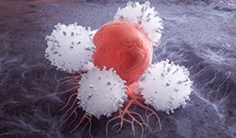
NK cells eliminate target cells through multiple mechanisms, including perforin/granzyme-mediated lysis; engagement of death receptor pathways, such as FasL and TRAIL; and antibody-dependent cellular cytotoxicity (ADCC) via CD16 (FcγRIIIa)-mediated recognition of antibody-coated targets.
ADCC is a key mechanism that contributes to the clinical activity of many therapeutic antibodies. This highlights the importance of NK cell–based functional assays for evaluating antibody efficacy during drug development.
Evaluate how antibody isotype, Fc mutations, and glycoengineering (afucosylation) affect NK cell–mediated killing with approved antibody benchmarking
Assess multispecific antibodies that bridge NK cells to tumor targets via CD16, NKp46, or other activating receptor engagement
Quantify killing activity of CAR-engineered NK cells for allogeneic “off-the-shelf” cellular therapy potency testing
Assess how checkpoint inhibitors, cytokines, or small molecules enhance NK cell killing when combined with therapeutic antibodies
Choose the readout best suited to your program: impedance for kinetics, flow cytometry for mechanistic depth, luminescence for throughput.
All ADCC assays validated using approved therapeutic antibodies (Trastuzumab, Rituximab, Cetuximab), ensuring clinically relevant benchmarking.
Routinely test across 3+ donors to characterize biological variability and identify consistent activity profiles across NK cell preparations.
Combine killing data with NK cell activation markers, receptor profiling, and cytokine secretion for comprehensive characterization.
Advance promising candidates to humanized xenograft models with NK cell reconstitution for in vivo ADCC or NK engager evaluation.
In this study example, we explore the effect of the monoclonal anti-Her2 antibody Trastuzumab (Trz) on NK cell killing by co-cultivating PBMCs with tumor cells expressing the cognate antigen.

Luciferase-expressing tumor cells exhibiting Her2 are seeded in a 384 well plate. PBMCs at different E:T (effector : target cell) ratios are added on top of the tumor cells. After an incubation period of 16 hours in the presence of Trastuzumab, the luciferase activity is measured as a readout of tumor cell viability.
Trastuzumab (Trz)-induced immune killing of luciferase-labeled SKOV-3 tumor cells.
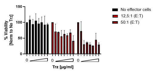
The bar graph shows the mean and SD of % viability normalized to the untreated (no Trz) condition.
In this study example, we explore the effect of a monoclonal antibody on NK cell killing by co-cultivating NK cells with tumor cells expressing the cognate antigen. Viability testing of the tumor cells was performed via flow cytometry.

NK cells were isolated from healthy donor PBMCs with a human NK cell isolation kit. Target cells were co-cultivated at different ratios for 4 hours in the presence of a monoclonal antibody recognizing a target cell-specific antigen. Tumor cells were detected via CellTracker Green dye and viable cells were quantified using a viability dye by flow cytometry.
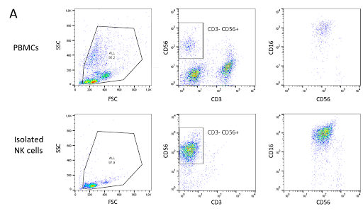
NK cells were isolated from healthy donor PBMCs via an NK Cell Isolation Kit. The purity of isolated NK cells was quantified by staining with NK cell-specific markers CD3-, CD56+, CD16+ before isolation (upper row) and after isolation (lower row). 10 million NK cells could be recovered from 300 million PBMCs with 88% purity.
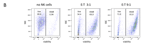
Viable target cells were quantified after a 4-hour co-cultivation of isolated NK cells and target cells in the presence of a target cell-specific antibody. The target cells were stained with CellTracker Green and subjected to viability testing with a live/dead dye via flow cytometry.
The presence of NK cells in a ratio of 3:1 increased the number of dead target cells from 9% to 65%. A ratio of 9:1 resulted in 86% of dead target cells.
Natural killer cell engagers are designed to tether NK cells to tumor cells. These multi-specific antibodies contain fragments specifically binding to NK cells and to tumor-associated antigens facilitating tumor cell killing.

NK cells were isolated from healthy donor PBMCs with a human NK cell isolation kit. The luciferase-expressing tumor target cells were co-cultivated with the NK cells at different ratios overnight in the presence of a bi-specific antibody recognizing NK cells and a tumor cell-specific antigen. At the end of the incubation period, the cells were lysed, and luciferase was quantified as a readout of tumor cell viability.
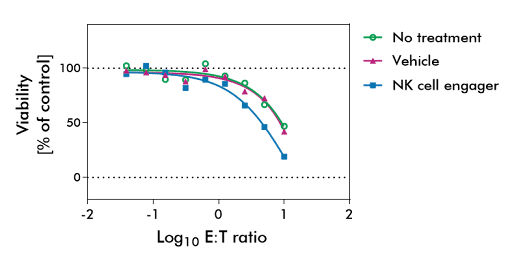
Co-cultivating NK cells and tumor target cells resulted in a decline of tumor cell viability in the presence and absence of the NK cell engaging test substance due to the innate function of NK cells to recognize and kill tumor cells. In the presence of a test substance, however, the tumor cell killing could be augmented significantly. At an effector:target cell ratio of 10:1, only 35% of tumor cells were viable in the presence of the NK cell engager compared to around 50% viable tumor cells in the control groups.
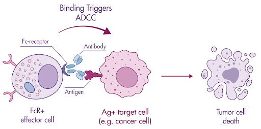
Components of the cell-mediated immune system target and kill virus-infected or other diseased (e.g., cancer) cells. When FcR-bearing effector cells identify a target cell that has been opsonized by antibodies, the ADCC pathway is activated. Once the effector cells have been activated by binding, the target cells will be killed by the cell-mediated ADCC immune defense mechanism.
Antibody-dependent cell-mediated target killing of virus-infected or other diseased cells.
Effector cells (Jurkat T cells) cultured for ADCC bioassays are engineered with stably expressing FcγRIIIa and have an NFAT response element that promotes the expression of firefly luciferase. Upon antibody binding to target antigens on the cell surface of the target cells (tumor cells, virus-infected cells, or other diseased cells), the Fc portion of the target-bound antibody binds to the FcγRIIIa receptors on the cell surface of the effector cell. Through multiple cross-links, the binding ensures precise cell-to-cell recognition, which ultimately results in ADCC activation. The result of activating this pathway leads to the death of the target cells.
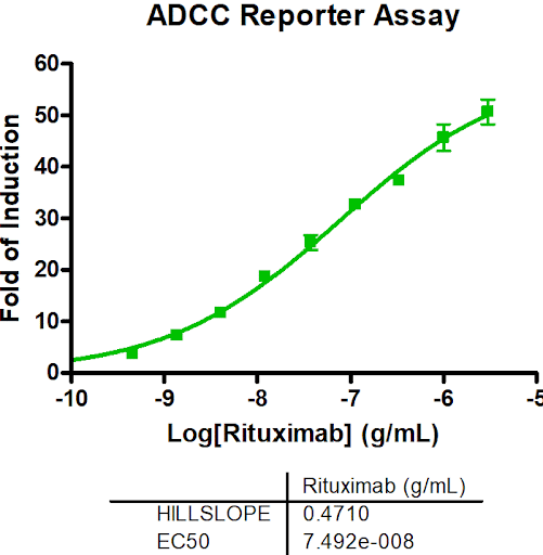
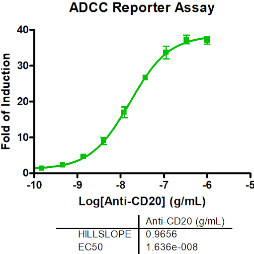
ADCC Reporter Bioassay was used to measure the Fc effector function of Rituximab to demonstrate assay specificity.
Direct NK killing occurs through activating receptor engagement when targets display stress ligands or lack MHC-I. ADCC requires antibodies that bind tumor antigens; NK cells recognize the Fc region via CD16, triggering killing. We can measure both and isolate antibody-dependent effects.
The optimal E:T ratio depends on whether PBMCs or isolated NK cells are used. Isolated NK cells are typically tested between 3:1 and 10:1, whereas PBMC assays often require 10:1 to 50:1. Higher ratios increase overall killing but demand more effector cells, while lower ratios can be more sensitive for detecting therapeutic effects. We recommend evaluating several ratios during assay development.
Yes, we routinely test across multiple donors (minimum 3 recommended) and report donor-to-donor variability. This is critical for understanding the range of expected clinical responses.
Standard controls include isotype-matched antibodies, NK cells alone (direct killing baseline), and tumor cells alone (background viability). Reference antibodies with known ADCC activity may serve as positive controls.
Cryopreserved NK cells provide reproducible results but may require longer incubation (24 hours vs. 4-6 hours for fresh). We offer both options depending on study requirements.
Yes, we accept customer-supplied CAR-NK cells and design potency assays using appropriate tumor target lines. We can also provide target cells expressing specific antigens.
From ADCC screening to NK cell engager development, our team provides the functional data needed to advance your antibody or cellular therapy program.