In Vivo Hollow Fiber Model
In Vivo Pharmacology

In Vivo Hollow Fiber Model

The In Vivo Hollow Fiber Model is a drug screening assay bridging the gap between in vitro and in vivo testing of anti-cancer agents allowing for rapid answers regarding drug efficacy.

The method is based on growing three tumor cell lines into one mouse, allowing a fast and economic screening approach with the following advantages:

  • Use the Hollow Fiber Model to select the best drug candidates and tumor models for subsequent xenograft tumor testing with higher predictability than conventional assays.
  • Any human tumor cell line can be included in a study. We can provide tumor cell lines of our collection of 253 cell lines or use a custom cell line for testing.
  • The Hollow Fiber Model can be performed early in the drug discovery process with simultaneous evaluation of pharmacokinetic and pharmacodynamic parameters.

Efficacy testing with confidence

Including the In Vivo Hollow Fiber Model in the drug discovery process allows for lead candidate identification and lead optimization before entering the phase of time-consuming human xenograft tumor testing. Better drugs are made faster and more cost-effective.

undefined

Study Details

  • Study Layout
  • Small compound study example
  • Antibody-treatment Example
Study Layout

Compounds
The fibers’ semipermeable membrane allows the crossing of test agents that are smaller than 500 kDa, such as small molecules or antibodies. Larger test items such as nanoparticles or cell-based therapies cannot be tested.

Standard study layout
Cells of three tumor cell lines are grown in cell culture and loaded each into a separate hollow fiber. The three fibers each loaded with one tumor cell line are cut into pieces and two pieces are implanted intraperitoneally and subcutaneously, respectively, into each mouse. Thus a total of 6 fibers is implanted into each mouse. After a drug treatment period of 14 days, the hollow fibers are harvested and analyzed for cell viability using CellTiter-Glo assay.

The protocol was adapted from Hollingshead et al., 1995, Life Sciences 57, pp. 131-141.

Report
A comprehensive report is prepared by a PhD-level medical writer. The report will be custom-tailored for each project with data that can be used for filing official documents. The report includes material, methods, raw data, animal health charts, and graphs plus statistical evaluation.

Add ons

  • During the, study blood samples can be taken from the mice to perform compound quantification
  • Depending on the study goal, fibers may be used for subsequent readouts such as protein or RNA quantification
Small compound study example

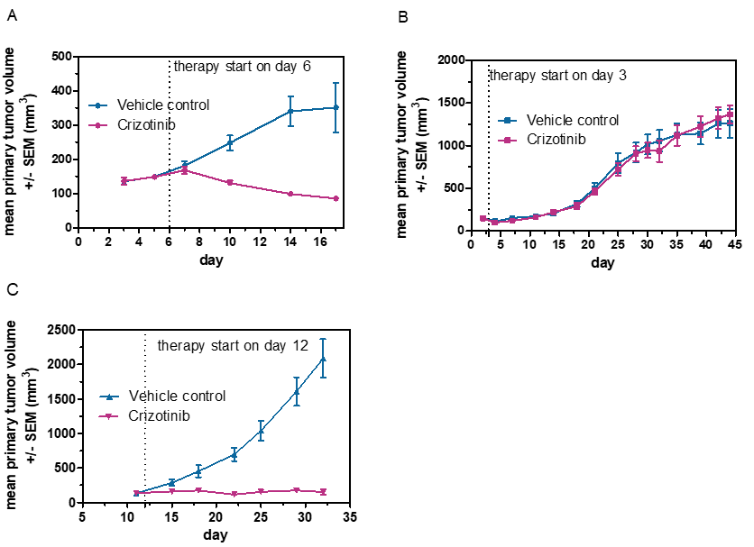
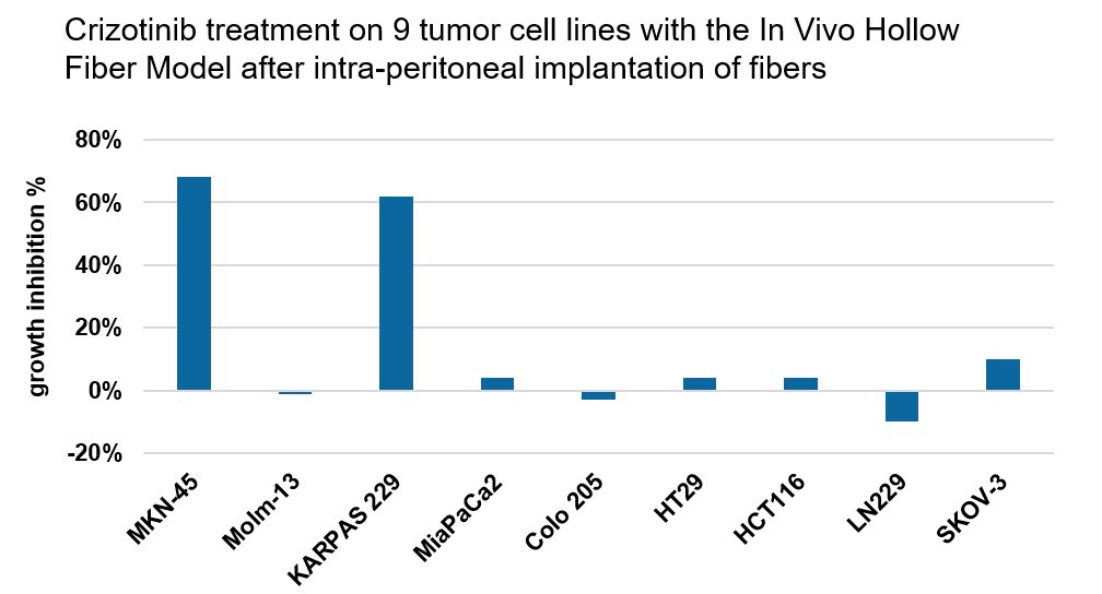
In the first part of a study, we determined the efficacy of a MEK kinase inhibitor on nine tumor cell lines in the In Vivo Hollow Fiber Model (treatment 50 mg/kg for two weeks). The mean of intraperitoneally implanted hollow fibers is shown as the ratio of treated versus untreated group (above).

In the second part, we tested the efficacy of the drug on MKN-45 (A), MiaPaCa2 (B), and KAPRAS-229 (C) in subcutaneous xenograft tumor models. In each case, the In Vivo Hollow Fiber Model was predictive for the drug’s efficacy on the subcutaneous xenograft tumor model.

Antibody-treatment Example

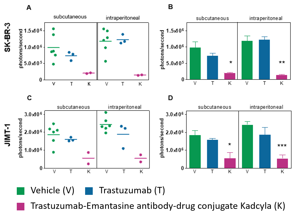
The efficacy of an antibody-drug conjugate was tested on two breast cancer cell lines with the Hollow Fiber Model.

Hollow fibers loaded with Her2 positive breast cancer cell lines SK-BR-3 and JIMT-1 were implanted in mice subcutaneously and intraperitoneally. The mice were treated with either vehicle (V), the antibody (T) or the andibody-drug conjugate (K) for two weeks. Hollow fibers were isolated and viable cells were quantified at end point of study.
Data are shown as single points (A, C with mean) or bar graphs (B, D with mean + SEM). ANOVA test was used to determine statistical significant results. * p ≤ 0.05, *** p ≤ 0.001

Result: None of the cell lines are inhibited by the treatment with the antibody because the drug acts via antibody-dependent cell-mediated cytotoxicity (ADCC), which is prevented in the Hollow Fiber Model because immune cells cannot enter the fibers.
In contrast, the antibody-drug conjugate acts directly on the cells via the cytotoxic agent bound to the antibody. This study shows that the Hollow Fiber Model is suitable for investigation of antibody-drug conjugates.