Cell-based Epigenetic Assays
Cell-Based Assays

Cell-based Epigenetic Assays

Cell-based epigenetic assays are valuable to probe new drug candidates in the physiologic environment of intact cells. They are the natural second step after the initial screening of new compounds in biochemical assays adding valuable biologically relevant information for the efficacy of the compounds.

Reaction Biology offers three options for cellular epigenetic inhibitor testing:

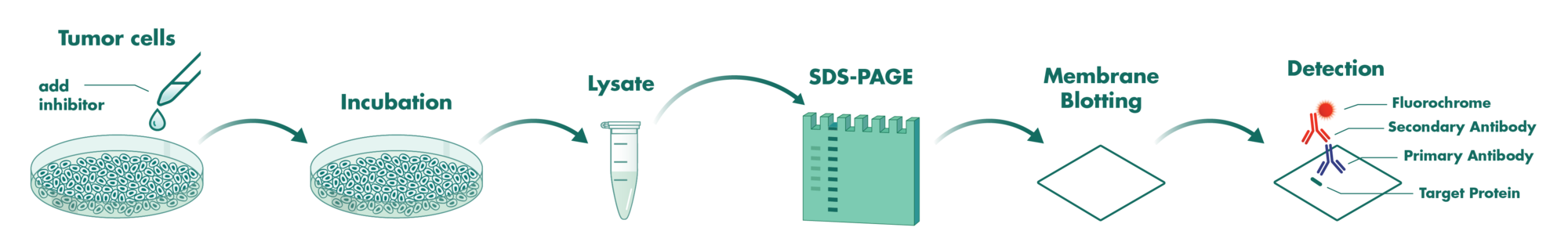
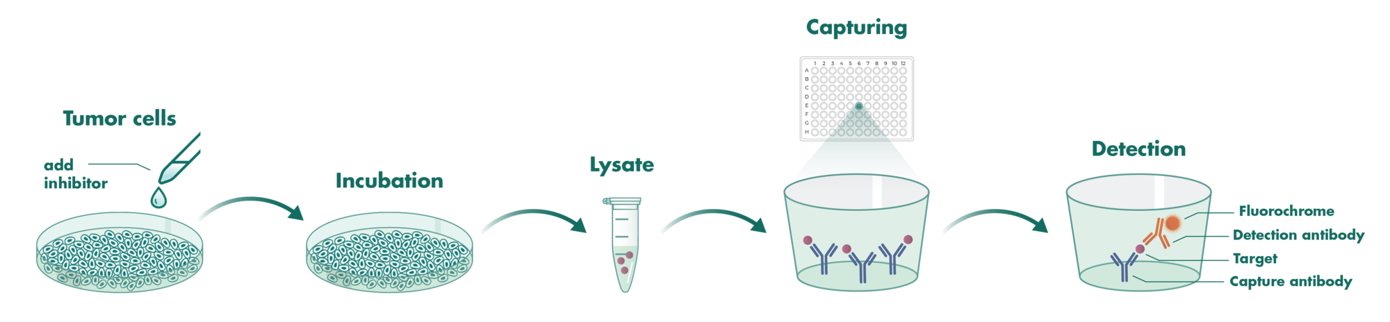
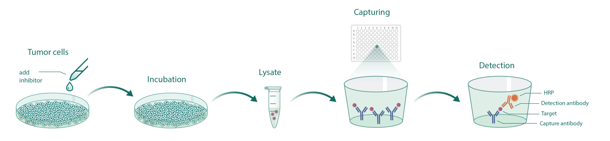
1. Functional Assays: Testing the modulation of the histones including acetylation, methylation, and phosphorylation.

2. Binding Affinity: Using Promega’s NanoBRET technologies we can test the binding affinity and residence time of new epigenetic compounds in intact cells

3. Protein:Protein Interaction: Testing the interaction of bromodomains with histones in intact cells based on Promega’s NanoBRET Bromodomain/Histone Interaction Assay.

Custom Cellular Epigenetic Assays

Cell-based Epigenetic Assay Details

  • Tubulin Deacetylation Assay
  • Histone Deacetylation Assay
  • Histone Methylation Assay
  • Histone Phosphorylation Assay
  • NanoBRET Bromodomain:Histone Interaction Assay
  • NanoBRET Target Engagement Epigenetic Assay
Tubulin Deacetylation Assay

Purpose: Quantification of acetylated tubulin as a readout of HDAC activity for drug screening

Targets: HDAC

Assay format: Western Blot


Cells incubate with the compound for 16 hours. Subsequently, cells are lysed, and the proteins are solubilized via a detergent from the cells. Next, the proteins are separated by their molecular size via SDS-PAGE before being transferred onto a membrane. Acetylated alpha-tubulin is detected by a specific antibody labeled with fluorochromes and scanned. The membrane is then reprobed with an antibody detecting tubulin for normalization purposes.
The membrane is scanned with the LICOR Odyssey Infrared imager for quantification of alpha-tubulin and acetylated alpha-tubulin in the sample.


Assay Format: ELISA

Cells incubate with the compound. Subsequently, cells are lysed, and the proteins are solubilized via a detergent. Next, the solution is transferred to an ELISA plate coated with anti-alpha-tubulin antibodies for capturing alpha-tubulin. After washing, an acetyl-lysine antibody is added to bind to acetylated alpha-tubulin. A secondary HRP-linked antibody is used for the quantification of bound detection antibodies which is proportional to the amount of acetylated alpha-tubulin in the sample.

Histone Deacetylation Assay

Purpose: Quantification of acetylated histones as a readout of HDAC activity for drug screening

Targets: HDAC

Assay format: ELISA

Cells incubate with the compound. Subsequently, cells are lysed, and the proteins are solubilized via a detergent. Next, the solution is transferred to an ELISA plate coated with anti-Histone H3 antibodies to capture Histone H3. After washing, an acetyl-lysine antibody is added to bind to acetylated Histone H3. A secondary HRP-linked antibody is used for quantification of bound detection antibodies which is proportional to the amount of acetylated Histone H3 in the sample.

Assay format: HDAC-Glo
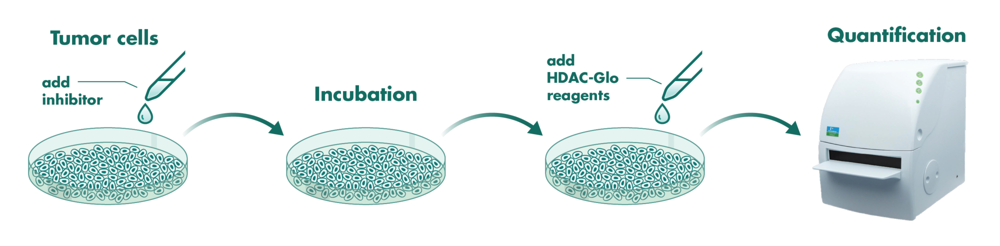
Cells incubate with the compound for 1 hour in serum-free medium. The HDAC-Glo substrate is added with is an acetylated peptide fused to aminoluciferin. After deacetylation of the peptide by HDAC activities, the peptide is cleaved via a protease in the reagent to release aminoluciferin substrate which is quantified in a reaction using luciferase. The luminescence is quantified via an Envision 2104 Multilabel Reader.

Histone Methylation Assay

Purpose: Quantification of methylated histone as a readout of methyltransferase activity for drug screening

Targets: Methyltransferase

Assay format: Western Blot

Cells incubate with the compound. Subsequently, cells are lysed, and the proteins are solubilized via a detergent. Next, the proteins are separated by their molecular size via SDS-PAGE before being transferred onto a membrane. Tri-methylated Histone H3 is detected by a specific antibody labeled with fluorochromes and scanned. The membrane is then reprobed with an antibody detecting Histone H3 for normalization purposes.

The membrane is scanned with the LICOR Odyssey Infrared imager for quantification of Histone H3 and tri-methylated Histone H3 in the sample.

Histone Phosphorylation Assay

Purpose: Quantification of phosphorylated histone as a readout of methyltransferase activity for drug screening

Targets: Kinases

Assay format: ELISA

Cells incubate with the compound for 90 minutes in serum-free media. After treatment with phosphatase inhibitors, cells are lysed, and the proteins are solubilized via a detergent. Next, the solution is transferred to an ELISA plate coated with anti-Histone H3 antibodies to capture Histone H3. After washing, a phospho-Histone 3 antibody is added to detect phosphorylated Histone H3. A secondary HRP-linked antibody is used for quantification of bound detection antibodies which is proportional to the amount of phosphorylated Histone H3 in the sample.

NanoBRET Bromodomain:Histone Interaction Assay

Purpose: Testing the efficacy of epigenetic drug candidates to interrupt the interaction of a bromodomain protein with chromatin

Targets: Bromodomains

Assay Format: NanoBRET Bromodomain:Histone Interaction Assay (Promega)


HEK 293T cells are transfected with two constructs: a bromodomain-NanoLuc fusion construct and a Histone-HaloTag fusion construct.

After 24 hours, the Halo Ligand and the test compound will be added. The interaction of bromodomain and histone will allow for electron transfer between the NanoLuc to the HaloLigand eliciting fluorescence in the presence of the NanoLuc substrate.

NanoBRET Target Engagement Epigenetic Assay

Purpose: Testing the binding affinity of epigenetic drug candidates to their target in intact cells

Targets: Bromodomains, HDAC

Assay Format: NanoBRET Intracellular Target Engagement Assay (Promega)
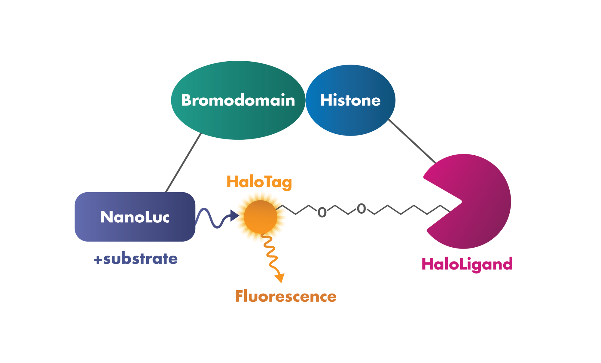
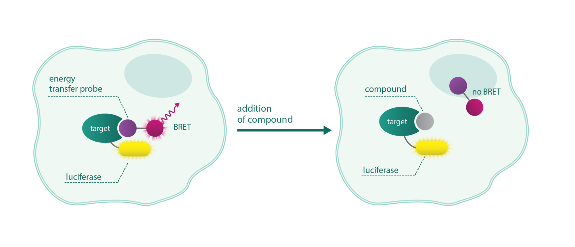
NanoBRET assays are compound competition assays that rely on bioluminescence resonance energy transfer (BRET) between a luciferase-tagged target and a fluorescent tracer. In the presence of the compound, the tracer is replaced diminishing the fluorescence.

Quantification and specificity are key attributes of the NanoBRET system.