BaF3 Cell Proliferation Assay
Target-specific Assays

BaF3 Cell Proliferation Assay

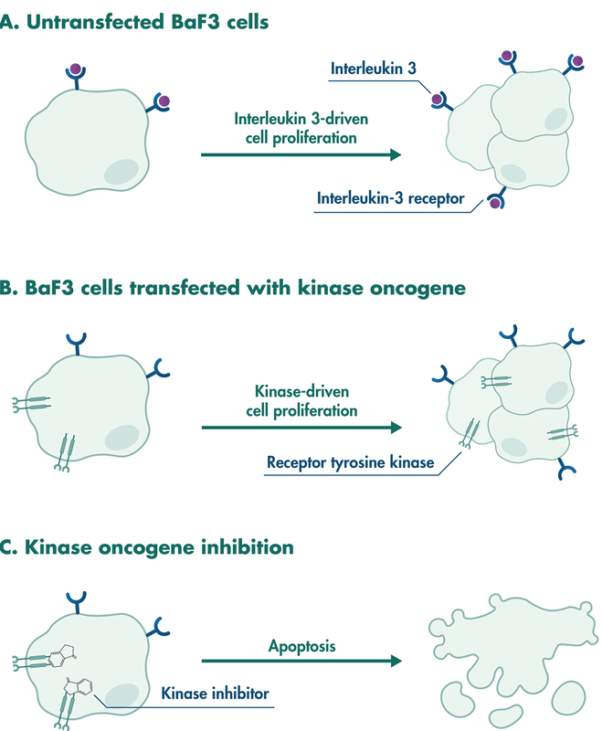
Reaction Biology performs compound testing with BaF3 cell lines stably expressing an oncogenic driver kinase. The principle of this assay is based on BaF3 cells, a pro-B-cell line that is dependent on interleukin 3 for its survival and proliferation. Transgenic overexpression of oncogenic kinases can transform the cell line to become independent of interleukin-3. Instead, cell proliferation is now dependent on the kinase signaling.

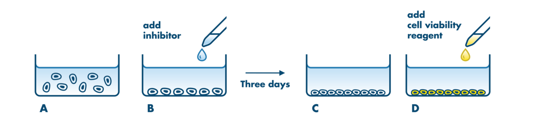
The assay is suitable to investigate if a compound is able to inhibit the activity of the transforming target kinase and – as a result – prevent the proliferation of these kinase-expressing BaF3 cells.

BaF3 Cell Proliferation Assays are helpful to answer a variety of research questions, including:

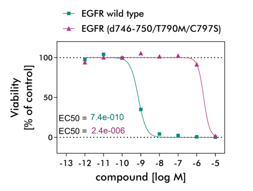
  • determination of the potency of kinase inhibitors
  • accessing the transforming capacity of new kinase mutations or kinase fusion proteins
  • predicting the resistance to small-molecule kinase inhibitors elicited by point mutations
  • investigating signaling events downstream of the kinase of interest
  • off-target screening via rescue experiments by the addition of interleukin-3

The BaF3 Cell Proliferation Assay is one of three cell-based assay formats offered for compound testing at Reaction Biology including the NanoBRET Intracellular Target Engagement Assay and the Cellular Phosphorylation Assay. All assays provide compound testing in the physiologic environment of intact cells providing biologically relevant and reproducibly data.

Find an overview of our cell-based assay formats for kinase screening here.

BaF3 Assay Principle

The overexpression of receptor tyrosine kinases enables the interleukin 3-dependent Ba/F3 cell line to grow without the supplement of interleukin 3. The inhibition of the activity of the transforming kinase leads to the loss of growth stimuli resulting in cell apoptosis.

Example cell lines for the BaF3 Proliferation Assay

BaF3 Cell Proliferation Assay Details

  • Readout
  • Assay Development
  • Assay Details
  • Study example
Readout

We determine the rate of cell proliferation via Cell Titre Glo, which quantifies the amount of ATP in live cells. Further readouts such as Alamar Blue, MTT, CyQuant, or IncuCyte Real-Time Imaging are options.

BaF3 cells stably expressing the kinase of interest are grown in multi-well plates without interleukin 3 (A). The kinase inhibitor is added and incubated for a 72 hour time period (B, C). The number of viable cells is determined by luminescence readout after adding CellTiter-Glo dye (D).

Assay Development

The BaF3 Proliferation Assay can be performed with constructs of receptor tyrosine kinases, in particular those enabling survival, growth, and cell proliferation.

A typical assay development project comprises:

  • molecular biology to create the plasmid
  • lentiviral cell line transduction
  • analysis of transgene expression
  • testing of cell proliferation with and without the addition of interleukin 3
  • validation via IC50 value determination of known kinase inhibitors in two independent projects

Asset: In addition to the investigation of the cell proliferation rate of the transformed cells, we may be able to set up a companion Cellular Phosphorylation Assay to determine the effects of kinase inhibition on substrate phosphorylation.

Assay Details

Setup: IC50 value determination and single concentration testing are an option. IC50 value determination is performed with 8 concentrations in duplicate.
This testing system is suited for low, medium, and high-throughput screening.

Controls: DMSO-only treatment serves as high control. Staurosporine treatment (at 1×10⁻⁵M) serves as low control. In every project, the IC50 value determination of a standard reference compound is included.

Report: A detailed report including assay conditions, methodology, graphical blots, and comprehensive evaluation of data as well as raw data for each analysis will be provided.

The turnaround time for standard studies is about 3 weeks. Expedited scheduling and data delivery can be arranged prior to study commencement.

Compound Information: Please see our FAQ page for detailed sample requirements and shipping instructions for cell-based screening (German Facility).

Screening facility: Assay development and CellTiter-Glo readouts are performed at our German research site.

Study example

BaF3 cells stably expressing human EGFR wild type and EGFR mutant d746-750/T790M/C797S were treated with the EGFR inhibitor Afatinib to determine the potency of the drug to disrupt EGFR cell signaling.

Cells were cultured in a medium supplemented with FCS in the absence of interleukin 3 and incubated overnight before compounds were added. After incubation for 72 h, CellTiterGlo reagent (Promega) was added and luminescence was measured with the Envision Multilabel Plate Reader.

The high and low values for IC50 value determination were DMSO-only high control and Staurosporin treatment low control.