ELISA (absorbance)
Sandwich capture and phospho-detection read on standard absorbance plate readers. It pairs a target-specific capture antibody with a phospho-specific detection antibody, the robust default format across the panel.

Cellular kinase inhibition quantified through substrate phosphorylation assays.
Percent inhibition and IC50 on physiological substrates across 51 validated target-specific models, wild type and mutant, with new targets built to order.
Kinases signal by transferring phosphate onto substrate proteins, and a large fraction of the proteome carries phosphorylation at any moment. A compound that blocks a kinase in a purified biochemical assay does not always block it inside a cell, where membrane permeability, physiological ATP, competing pathways, and target expression all shape the result. The cellular phosphorylation assay measures the event that matters therapeutically: inhibition of substrate phosphorylation by the intended kinase, in intact cells, reported as percent inhibition and IC50.
Sandwich capture and phospho-detection read on standard absorbance plate readers. It pairs a target-specific capture antibody with a phospho-specific detection antibody, the robust default format across the panel.
Time-resolved, bi-antibody FRET detection. Time-resolved reading suppresses short-lived medium autofluorescence for a high signal-to-noise ratio.
Homogeneous, no-wash bead-proximity readout. Donor and acceptor beads emit signal only when the captured target and its phospho-epitope are held in proximity, which lowers handling and background.
Receptor tyrosine kinases are read by pan-phospho-tyrosine antibodies on the receptor itself; cytosolic kinases are read on a direct substrate protein. Examples include ERK1/2 for MEK1/2, GSK3-beta Ser9 for AKT1, MEK1 Ser218/222 for B-RAF V600E, and p53 Ser15 for ATM.
| Resistance-mutant potency | EGFR and MET are built as wild type and resistance mutants in a shared background, so a compound’s potency shift across the mutant series is read directly at the level of cellular catalytic activity. The MET series spans D1228N, F1200I, and the Y1230 cluster; the EGFR series spans the classical activating, T790M, and C797S resistance combinations. |
| Substrate-based readouts | Receptor kinases report their own activity through autophosphorylation, read directly on the captured receptor with pan-phospho-tyrosine antibodies. Cytosolic kinases are read on a direct substrate one step down the pathway:
Each is anchored to a target-specific reference inhibitor. |
| Plasma inhibitory assay | Cellular potency is re-measured in the presence of plasma to quantify the effect of plasma protein binding on a compound. This can be run in vitro with plasma-supplemented medium or ex vivo using blood drawn from animals after in vivo dosing, linking cellular IC50 to real exposure. |
| Custom model development | Targets, mutants, and downstream-substrate readouts outside the catalog are generated in the same validated workflow, including wild-type-versus-mutant pairs in a matched background. This is the most common entry point for programs whose target of interest is not yet a standard model. |

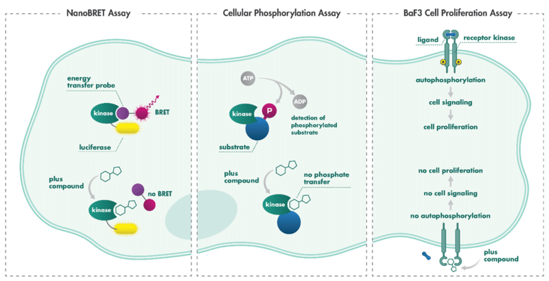
Reaction Biology measures cellular kinase activity by using phospho-specific antibodies to quantify substrate phosphorylation, identifying when a drug candidate successfully blocks enzyme function. A reduction in signal indicates that a compound has crossed the cell membrane and inhibited the targeted kinase.
The assay quantifies catalytic output on a physiological substrate in living cells, the functional consequence of inhibition. It complements NanoBRET target engagement, which reports binding, by confirming that binding translates into blocked signaling.
Permeability, physiological ATP, target expression, and competing pathways are all in play, so cellular IC50 values carry information a purified biochemical assay cannot provide.
Selected targets, including EGFR and MET, are built as wild type and resistance mutants in the same cellular background, giving clean potency shifts at the level of cellular catalytic activity.
Every model is benchmarked against a known inhibitor of that kinase, delivering reproducible IC50 values across the validated panel.
The impact of plasma protein binding on cellular potency can be measured, either ex vivo using blood from dosed animals or in vitro by supplementing the culture medium with plasma, to connect biochemical potency to exposure.
When your target or specific mutant is not in the catalog, a new model is generated inside the same validated framework, including new downstream-substrate readouts where a direct substrate is unavailable.
ELISA, AlphaLISA, and HTRF are selected per target-specific substrate to give the cleanest, most quantitative phospho-signal.
It is a cell-based kinase activity assay. In intact cells, the target kinase is activated and the resulting phosphorylation of a physiological substrate is quantified, so a compound’s ability to inhibit that kinase inside a living cell is reported as percent inhibition and IC50.
A biochemical assay measures phosphoryl transfer with purified enzyme and substrate. The cellular phosphorylation assay measures the same catalytic output inside a living cell, where permeability, physiological ATP, target expression, and competing pathways all influence the result. It is typically used to confirm that biochemical potency carries into a cellular setting.
NanoBRET measures compound binding to the kinase in cells. The cellular phosphorylation assay measures kinase activity, the functional consequence of that binding. Programs often run NanoBRET for binding breadth, then a cellular phosphorylation assay for activity and potency depth.
Percent inhibition of cellular kinase activity and IC50, generated from 8 compound concentrations in duplicate, with the phospho-substrate signal quantified by sandwich immunoassay.
ELISA (absorbance), AlphaLISA (fluorescence), and HTRF (fluorescence, bi-antibody), selected per target. The sandwich principle, one target-specific antibody plus one phospho-specific antibody, requires two independent epitopes and increases selectivity.
Yes. Selected targets, including EGFR and MET, are built as wild type and resistance mutants in the same cellular background for direct potency comparison. New mutants can be generated on request.
Yes. New target-specific models, mutants, and downstream-substrate readouts are built to order in the same validated framework. Most inbound programs begin here.
A version of the cellular phosphorylation assay run in the presence of plasma to quantify how plasma protein binding affects a compound’s cellular potency, either in vitro with plasma-supplemented medium or ex vivo from dosed-animal blood.
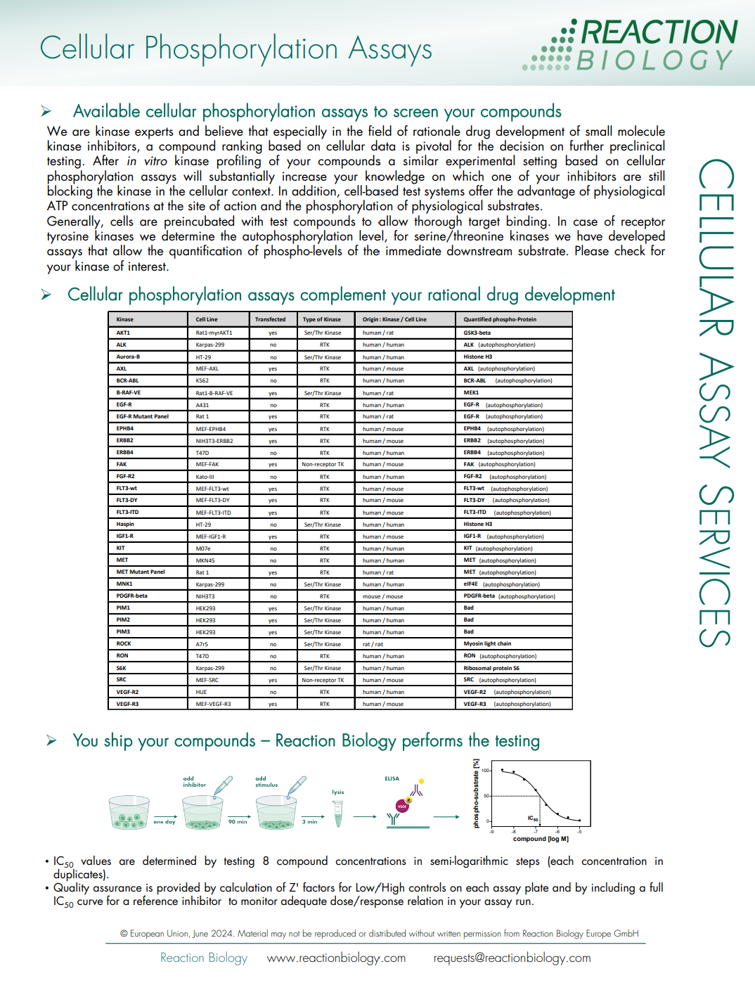
| Target | HGNC name | UniProt | Kinase class | Type | Cell Line | Expression | Info sheet |
|---|---|---|---|---|---|---|---|
| AKT1 | AKT1 | P31749 | Serine/threonine kinase | Wild type | Rat1 | Transfected | Data sheet |
| ALK | ALK | Q9UM73 | Receptor tyrosine kinase | Wild type | Karpas-299 | Endogenous | Data sheet |
| ATM | ATM | Q13315 | Serine/threonine kinase | Wild type | U2OS | Endogenous | Please inquire |
| Aurora B | AURKB | Q96GD4 | Serine/threonine kinase | Wild type | HT-29 | Endogenous | Data sheet |
| AXL | AXL | P30530 | Receptor tyrosine kinase | Wild type | MEF | Transfected | Data sheet |
| BCR-ABL | BCR-ABL1 | P11274 / P00519 | Cytoplasmic tyrosine kinase | Fusion | K562 | Endogenous | Data sheet |
| BRAF V600E | BRAF (V600E) | P15056 | Serine/threonine kinase | Mutant | Rat1 | Transfected | Data sheet |
| c-KIT | KIT | P10721 | Receptor tyrosine kinase | Wild type | M07e | Endogenous | Data sheet |
| c-MET | MET | P08581 | Receptor tyrosine kinase | Wild type | MKN45 | Endogenous | Data sheet |
| c-MET (D1228N) | MET (D1228N) | P08581 | Receptor tyrosine kinase | Mutant | Rat1 | Transfected | Data sheet |
| c-MET (D1288H) | MET (D1288H) | P08581 | Receptor tyrosine kinase | Mutant | Rat1 | Transfected | Data sheet |
| c-MET (F1200I) | MET (F1200I) | P08581 | Receptor tyrosine kinase | Mutant | Rat1 | Transfected | Data sheet |
| c-MET (Y1230A) | MET (Y1230A) | P08581 | Receptor tyrosine kinase | Mutant | Rat1 | Transfected | Data sheet |
| c-MET (Y1230D) | MET (Y1230D) | P08581 | Receptor tyrosine kinase | Mutant | Rat1 | Transfected | Data sheet |
| c-MET (Y1230H) | MET (Y1230H) | P08581 | Receptor tyrosine kinase | Mutant | Rat1 | Transfected | Data sheet |
| c-SRC | SRC | P12931 | Cytoplasmic tyrosine kinase | Wild type | MEF | Transfected | Data sheet |
| EGFR | EGFR | P00533 | Receptor tyrosine kinase | Wild type | A431 | Endogenous | Data sheet |
| EGFR (d746-750/C797S) | EGFR (d746-750/C797S) | P00533 | Receptor tyrosine kinase | Mutant | Rat1 | Transfected | Data sheet |
| EGFR (d746-750/T790M) | EGFR (d746-750/T790M) | P00533 | Receptor tyrosine kinase | Mutant | Rat1 | Transfected | Data sheet |
| EGFR (d746-750/T790M/C797S) | EGFR (d746-750/T790M/C797S) | P00533 | Receptor tyrosine kinase | Mutant | Rat1 | Transfected | Data sheet |
| EGFR (d747-749/A750P) | EGFR (d747-749/A750P) | P00533 | Receptor tyrosine kinase | Mutant | Rat1 | Transfected | Data sheet |
| EGFR (d752-759) | EGFR (d752-759) | P00533 | Receptor tyrosine kinase | Mutant | Rat1 | Transfected | Data sheet |
| EGFR (G719S) | EGFR (G719S) | P00533 | Receptor tyrosine kinase | Mutant | Rat1 | Transfected | Data sheet |
| EGFR (L858R) | EGFR (L858R) | P00533 | Receptor tyrosine kinase | Mutant | Rat1 | Transfected | Data sheet |
| EGFR (L858R/T790M) | EGFR (T790M/L858R) | P00533 | Receptor tyrosine kinase | Mutant | Rat1 | Transfected | Data sheet |
| EGFR (L861Q) | EGFR (L861Q) | P00533 | Receptor tyrosine kinase | Mutant | Rat1 | Transfected | Data sheet |
| EGFR (T790M) | EGFR (T790M) | P00533 | Receptor tyrosine kinase | Mutant | Rat1 | Transfected | Data sheet |
| EGFR (T790M/C797S/L858R) | EGFR (T790M/C797S/L858R) | P00533 | Receptor tyrosine kinase | Mutant | Rat1 | Transfected | Data sheet |
| EGFR (wild type control) | EGFR | P00533 | Receptor tyrosine kinase | Reference control | Rat1 | Transfected | Data sheet |
| EPHB4 | EPHB4 | P54760 | Receptor tyrosine kinase | Wild type | MEF | Transfected | Data sheet |
| ERBB2/HER2 | ERBB2 | P04626 | Receptor tyrosine kinase | Wild type | NIH3T3 | Transfected | Data sheet |
| ERBB4/HER4 | ERBB4 | Q15303 | Receptor tyrosine kinase | Wild type | T47D | Endogenous | Data sheet |
| FAK/PTK2 | PTK2 | Q05397 | Cytoplasmic tyrosine kinase | Wild type | MEF | Transfected | Data sheet |
| FGFR2 | FGFR2 | P21802 | Receptor tyrosine kinase | Wild type | Kato III | Endogenous | Data sheet |
| FLT3 (D835Y) | FLT3 (D835Y) | P36888 | Receptor tyrosine kinase | Mutant | MEF | Transfected | Data sheet |
| FLT3 (ITD) | FLT3 (ITD) | P36888 | Receptor tyrosine kinase | Mutant | MEF | Transfected | Data sheet |
| FLT3 (wild type control) | FLT3 | P36888 | Receptor tyrosine kinase | Reference control | MEF | Transfected | Data sheet |
| FLT4/VEGFR3 | FLT4 | P35916 | Receptor tyrosine kinase | Wild type | MEF | Transfected | Data sheet |
| FYN | FYN | P06241 | Cytoplasmic tyrosine kinase | Wild type | Rat1 | Transfected | Data sheet |
| HPK1/MAP4K1 | MAP4K1 | Q92918 | Serine/threonine kinase | Wild type | Jurkat | Endogenous | Data sheet |
| IGF1R | IGF1R | P08069 | Receptor tyrosine kinase | Wild type | MEF | Transfected | Data sheet |
| KDR/VEGFR2 | KDR | P35968 | Receptor tyrosine kinase | Wild type | HUE | Endogenous | Data sheet |
| LRRK2 | LRRK2 | Q5S007 | Serine/threonine kinase | Wild type | A549 | Endogenous | Data sheet |
| MEK1/2 | MAP2K1/MAP2K2 | Q02750 / P36507 | Dual-specificity kinase | Wild type | PANC-1 | Endogenous | Data sheet |
| MET (wild type control) | MET | P08581 | Receptor tyrosine kinase | Reference control | Rat1 | Transfected | Data sheet |
| PDGFRb | PDGFRB | P09619 | Receptor tyrosine kinase | Wild type | NIH3T3 | Endogenous | Data sheet |
| PIM1 | PIM1 | P11309 | Serine/threonine kinase | Wild type | HEK293T | Transfected | Data sheet |
| PIM2 | PIM2 | Q9P1W9 | Serine/threonine kinase | Wild type | HEK293T | Transfected | Data sheet |
| PIM3 | PIM3 | Q86V86 | Serine/threonine kinase | Wild type | HEK293T | Transfected | Data sheet |
| ROCK | ROCK1 and ROCK2 | Q13464 / O75116 | Serine/threonine kinase | Wild type | A7r5 | Endogenous | Data sheet |
| RON/MST1R | MST1R | Q04912 | Receptor tyrosine kinase | Wild type | T47D | Endogenous | Data sheet |