3D Tumor Spheroid Assay
Cell-Based Assays

3D Tumor Spheroid Assay Service

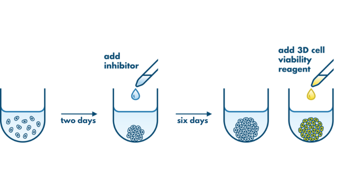
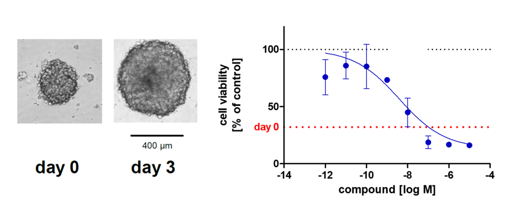
The 3D Tumor Spheroid Assay is used to analyze the therapeutic effect and tumor-penetrating capacity of anti-cancer therapeutics. This 3D assay is performed on spheroids with high translational value making it a valuable tool for lead candidate selection for in vivo studies.

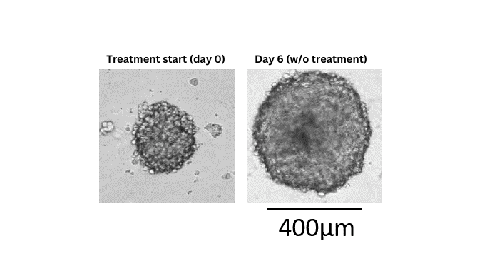
3D tumor spheroids recapitulate some of the complex processes that compounds face in an in vivo situation such as cellular barriers and cells adapted to gradients of oxygen and nutrient concentrations. The spheroids are composed of cells in various proliferative and metabolic states similar to tumors. Therefore, the 3D Tumor Spheroid Assay has a high predictive value for pre-clinical drug research.

The 3D Tumor Spheroid Assay is one of two 3D assays offered by Reaction Biology. The 3D Tumor Spheroid Assay is used for testing a drug’s anti-proliferative and cytotoxic capacity on existing tumor spheroids. In the Soft Agar Assay, which is also a 3D assay, we test the impact of drugs on the proliferation of single cells growing into colonies.

  • Mono-spheroid assay with human tumor cell lines
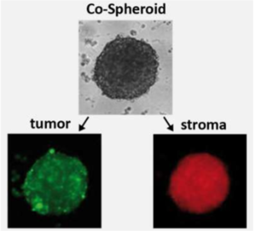
  • Co-culture spheroid testing with quantification of both, stroma and tumor cells
  • High-throughput compatible setup

3D Tumor Spheroids

Using spheroids for efficacy testing allows the detection of both cytotoxic and anti-proliferative effects of drugs.

Tumor cells assemble in ultra-low attachment multiwell plates to form spheroids. Within 6 days, they grow to a multiple of their original size allowing testing compounds for proliferation, or killing, of cells in the original spheroid. The example shown on the right: U87MG glioblastoma tumor spheroids at treatment start (day 0) and day 6 (w/o treatment).

U87 Monospheroid day 0 and day 6

U87 Monospheroid day 0 and day 6

3D Tumor Spheroid Assay Details

  • Procedure: Mono-Culture Spheroids
  • Mono-Culture Spheroids
  • Co-Culture Spheroids
  • Screening Formats
Procedure: Mono-Culture Spheroids

230623 monospheroid assay procedure

Assay procedure. Tumor cells are seeded in 384-well ultra-low attachment plates, shortly spun down and let to autonomously assemble spheroids. Compounds incubate for six days before readout.

Mono-Culture Spheroids

Mono-Culture Spheroids

Example of U87MG mono-spheroids. Left: Comparison of the size of U87MG spheroids at day 0 and 3. Right: Concentration-response curve of treatment with a small molecule MEK1/MEK2 inhibitor on U87MG mono-spheroids. Positive control (0 % viability) is staurosporine treatment; negative control (100 % viability) is vehicle control. ‘day 0’ shows the number of viable cells at the start of treatment.

Co-Culture Spheroids

3d tumor spheroids with luciferase to quantify co-culture

Assay procedure for co-culture spheroids: Firefly-luciferase expressing tumor cells and renilla-expressing stroma cells are seeded in a round-bottom 96 well plate and left to assemble for spheroid formation for three days. Compounds incubate for three days before firefly-luciferase and renilla-luciferase quantification from the lysate of the spheroid. The cytotoxic and anti-proliferative effects of compounds can be determined for both tumor and stroma cells separately.

3d tumor spheroids with luciferase to quantify co-culture

Example showing reduced sensitivity of DLD1 cells against a MEK kinase inhibitor in co-spheroids: DLD1 tumor cells were cultured in spheroids as mono-culture (black) or together with HS27A stroma cells (blue). Spheroids were treated for 72 h with the drug. Subsequently, the viability of tumor cells was analyzed by measurement of firefly luminescence.

Screening options:

  1. Co-spheroids with tumor & stroma cells, luciferase measurement only in tumor cells
  2. Co-spheroids with firefly luciferase-labeled tumor & renilla-luciferase labeled stroma cells
Screening Formats

Setups: IC50 value determination and single concentration testing are possible.

Cell lines: Please inquire to receive a list of cell lines that are established in the 3D Tumor Spheroid Assay format. New assays based on additional cell lines can be established according to the client’s research needs.

Controls: DMSO only treatment serves as high control. Staurosporine treatment (at 1×10⁻⁵M) serves as low control.

Turnaround time: Results will be available after 3 to 4 weeks. Expedited scheduling and data delivery can be arranged prior to study commencement.

Report: A detailed report including assay conditions, methodology, and comprehensive evaluation of data as well as raw data for each analysis will be provided. Photographs are optional.

Screening Facility: The assay is performed in Freiburg, Germany.

Compound requirements: In brief: 30 µl of the stock solution with 1000x of the highest assay concentration or the equivalent of solid material.