Browse the TMA Gallery
Access histology images of our syngeneic tumor models to make informed decisions about which model best fits your immunotherapeutic’s mechanism of action.

Visualize immune cell localization and target expression in tumor tissue
IHC reveals what flow cytometry cannot—where immune cells are located within tumor architecture and whether they penetrate into tumor tissue or remain confined to the stroma.
Flow cytometry quantifies which immune cells are present; immunohistochemistry shows you where they are. IHC enables visualization of tumor-infiltrating immune cells within tissue architecture—distinguishing stromal accumulation from true tumor infiltration. Our tissue microarray containing cores from syngeneic mouse models allows rapid comparison of immune infiltration patterns across tumor types, helping you select the optimal model for your mechanism of action before committing to efficacy studies.
Tissue microarrays consolidate dozens of tumor samples onto a single slide for standardized, high-throughput analysis:
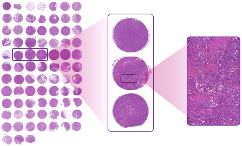
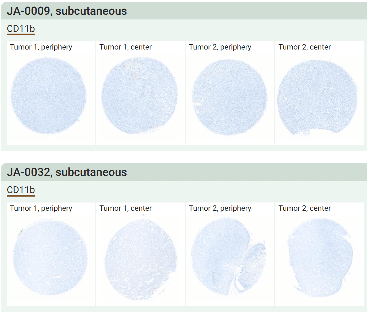
Browse histology and immune cell staining across our syngeneic tumor model portfolio. Images stained with H&E, anti-CD3, anti-CD4, anti-CD8, and anti-CD11b are available to inform your model selection before study initiation.
Access histology images of our syngeneic tumor models to make informed decisions about which model best fits your immunotherapeutic’s mechanism of action.
Request staining of our TMA with antibodies targeting your protein of interest, immune markers, or target antigens beyond our standard panel.
Receive unstained TMA slides for staining and analysis in your own laboratory using your established protocols.
We create custom TMAs from tumors harvested during your animal studies—enabling streamlined endpoint analysis across treatment groups on a single slide.
Visualize whether tumor-infiltrating immune cells remain in the stromal compartment or migrate into tumor parenchyma—critical context that flow cytometry cannot provide.
Multiplex IHC enables co-staining of multiple markers on a single section, revealing functional relationships between immune populations and answering specific mechanistic questions.
Quantify and localize target protein expression in tumor tissue to validate target engagement or confirm antigen expression in your model of choice.
IHC is typically performed when flow cytometry or MSD has yielded promising results, adding spatial context to quantitative immune profiling data.
| Marker | Target Population |
|---|---|
| H&E | General histology / tumor architecture |
| CD3 | Pan T cells |
| CD4 | Helper T cells |
| CD8 | Cytotoxic T cells |
| CD11b | Myeloid cells |
| Custom | Upon request |
Flow cytometry provides quantitative immune cell frequencies and multi-marker phenotyping with high throughput. IHC shows spatial localization within tissue architecture. We often recommend flow cytometry as the primary quantitative readout, with IHC as follow-up to visualize and confirm key findings.
Our TMA contains cores from our syngeneic mouse model portfolio. Browse the TMA Gallery to see available models and existing staining data for CD3, CD4, CD8, and CD11b.
Yes, multi-marker IHC can co-localize immune populations on a single section to answer specific functional questions about immunotherapeutic effects.
Yes, we can ship unstained TMA slides for in-house processing using your established protocols and antibodies.
Yes, de novo TMAs can be constructed from tumors harvested during your animal studies, enabling efficient endpoint analysis across all treatment groups on a single slide.
Our TMA is focused specifically on syngeneic mouse models for immuno-oncology research—enabling rapid model selection and IO-relevant endpoint analysis rather than broad biomarker discovery across human tumor banks.