Standard Efficacy Studies
Determine the therapeutic effect of a new drug on a syngeneic tumor model. Tumors are monitored by calipering after implantation; animals are randomized by tumor size before treatment begins.

Next-generation implantation for homogeneous growth and superior statistical power
A superior implantation method for syngeneic cell line-derived tumor models. Mammary fat pad injection overcomes ulceration, extending treatment windows and delivering reproducible study outcomes.
Standard subcutaneous implantation frequently causes tumor ulceration, forcing early study termination, shortening treatment windows, and introducing heterogeneity that undermines statistical power. SubQperior addresses these limitations through mammary fat pad implantation. The fat pad surrounds tumor cells with stroma and acts as a buffer zone between the tumor and dermis, restricting ulceration while supporting larger, more uniform tumor growth than conventional subcutaneous methods.
The SubQperior Advantage
Tumor cell implantation into the mammary fat pad allows growth in stroma-rich tissue. The fat pad restricts ulceration and supports superior tumor growth compared to subcutaneous implantation.
Determine the therapeutic effect of a new drug on a syngeneic tumor model. Tumors are monitored by calipering after implantation; animals are randomized by tumor size before treatment begins.
Determine suitable drug doses for efficacy testing across a defined concentration range.
Determine drug concentration in plasma and within tumor tissue in tumor-bearing mice.
Evaluate mechanism of action in vivo. Exploratory studies use flow cytometric analysis to investigate effects on tumor-infiltrating immune cells.
Assess synergistic effects of combination regimens. Our models are validated with immune checkpoint inhibitors anti-PD1, anti-PD-L1, and anti-CTLA-4, which are frequently used in combination therapy.
Elucidate whether and how a drug acts on its target, for example via flow cytometric analysis of tumor-infiltrating immune cells.
Tumor cells implanted into the mammary fat pad of the mouse strain.
When tumors reach predetermined volume, animals are randomized into treatment groups based on tumor size, and treatment begins.
Tumor size measured by caliper twice weekly, animal weight three times weekly, and behavior observed daily. Weekly graphical study updates provided.
Tumors isolated at necropsy for volume and wet weight. A PhD-level medical writer prepares a comprehensive report with raw data, methods, health charts, graphs, and statistical evaluation suitable for regulatory filing.
| Model | Cancer Type | Mouse Strain | anti-PD1 Response (TGI) | Immune Phenotype |
| MC38-CEA | Colon | C57BL/6 | High | Responsive |
| Clone M3 | Melanoma | DBA/2 | High | Responsive |
| CT26wt | Colon | BALB/c | Moderate-High | Responsive |
| HEPA 1-6 | Hepatoma | C57BL/6 | Moderate | Intermediate |
| EMT6 | Breast | BALB/c | Moderate | Intermediate |
| RENCA | Renal | BALB/c | Low-Moderate | Intermediate |
| LL-2 | Lung | C57BL/6 | Low | Low responder |
| 4T1 | Breast | BALB/c | Low | Low responder |
| B16-F10 | Melanoma | C57BL/6 | Variable | Cold tumor |
| AB12 | Mesothelioma | BALB/c | Low | Low responder |
Mammary fat pad implantation reduces ulceration, the leading cause of early study termination in subcutaneous models, extending usable treatment windows.
Homogeneous, reproducible growth combined with larger tumor sizes yields efficacy data with superior statistical power and fewer confounding dropouts.
Models characterized across a range of immune phenotypes and validated with anti-PD1, anti-PD-L1, and anti-CTLA-4 for combination study design.
Pair efficacy data with flow cytometry immune profiling, MSD cytokine quantification, histology, and PK/PD analysis within a single study.
Develop new models from customer-provided or commercially sourced cell lines. Establishment studies monitor growth characteristics across 12 mice. Cost-sharing options available.
Our animal facility in Freiburg, Germany is certified under ISO 9001:2015 for quality management. We operate under GV-SOLAS and ISO 9001 standards for animal welfare and code of practice.
All animal work conducted according to the 3R principles (Replacement, Reduction, Refinement). Three veterinarians and trained staff ensure the highest welfare standards, with regular inspection by regulation officers.
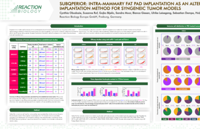
Goal: The frequently used immune checkpoint inhibitors anti-mPD-1 and anti-mCTLA-4 were tested for their potential to inhibit subQperiorTM CT26wt primary tumor growth.
Study layout: CT26wt cells were implanted into the mammary fat pad of Balb/C mice, at 12 mice per group. After randomization on day 5, treatments were initiated on the same day. Test compounds anti-mPD-1 (Group 2) and anti-mCTLA-4 (Group 3), both administered i.p. at 10.0 mg/kg on days 5, 8, and 12, were evaluated versus the corresponding vehicle control (Group 1). Tumor growth was monitored by calipering twice per week, animal weight was measured three times per week, animal behavior was observed daily. On day 21, animals were euthanized and endpoint measurements for the determination of primary tumor volumes, and wet weights were performed.

Animal weight of CT26wt subQperior tumor model is shown with vehicle and anti-mPD-1 and anti-mCTLA-4 treatment.


Tumor growth is shown for individual tumors. upper blot: anti-mPD-1 treatment vs vehicle treatment. lower blot: anti-mCtLA-4 treatment vs vehicle treatment.

At day 21, tumors were isolated from the mice at necropsy and weighed. Blotted are the mean tumor weights in the top graph and the individual tumor weights in the lower graph together with their median values and interquartile ranges. P values were calculated compared to the vehicle control using the unpaired t‑test and the one-way ANOVA with Dunnett´s test.
SubQperior is implantation of syngeneic tumor cells into the mammary fat pad rather than standard subcutaneous injection. The fat pad surrounds cells with stroma and buffers the tumor from the dermis, reducing ulceration and supporting larger, more homogeneous tumor growth.
More homogeneous and reliable tumor growth reduces variability between animals. This yields cleaner efficacy data, can reduce the number of mice required per arm, and minimizes dropouts from early ulceration-driven termination.
Validated models include CT26wt, MC38-CEA, Clone M3, B16-F10, 4T1, RENCA, EMT6, LL-2, and others spanning colon, breast, melanoma, renal, lung, and additional cancer types. Custom model development is available.
Yes. Most models have been characterized with anti-PD1, anti-PD-L1, and anti-CTLA-4, supporting both monotherapy benchmarking and combination study design.
Yes. Flow cytometry immune profiling is available as a study endpoint, including a >27-marker panel and specialized panels for TILs, and CNS tumor microglia.
Studies generally begin with tumor implantation 3 to 5 weeks after order receipt. You will receive weekly updates and direct contact with your study supervisor throughout.