TMA
Biomarker Discovery

TMA and Immunohistochemistry

Tissue microarrays (TMA) are effective tools for the histological analysis of tumors to discover biomarkers and characterize the mode of action of new drug candidates. They are also used for the analysis of target protein expression in a large set of tumors of different origins simultaneously to identify suitable mouse models for anticancer drug efficacy studies.

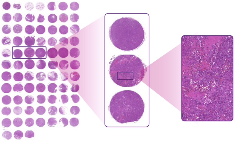
Reaction Biology’s TMA is a paraffin block that contains tumor cores from syngeneic mouse models available for efficacy testing at our in vivo pharmacology unit.

The tumor tissue microarray empowers to:

  • Select the best tumor model based on target expression
  • Ensure the immune population needed for your mode of action is present in the tumor model of your choice
  • Identify and measure biomarkers predicting the response to your drug candidate

Images of our tumors stained with H&E, anti-CD3, anti-CD11b, anti-CD4 and anti-CD8 antibodies can be viewed in our interactive TMA Gallery.

Contact us to request a custom stain of our TMA or receive slides of our TMA for in-house staining.

Caught in the act.

A TMA enables the comparison of various tumor models in regards to their histology, target expression, and immune cell infiltration.

Target proteins or immune markers are visualized on a cellular level understanding their location in stromal or tumoral tissue.

Enter our TMA Gallery to investigate the histology and immune cells of our syngeneic tumor models here.

tumor tissue microarray

Options to use Reaction Biology’s TMA for your cancer drug research project

Our TMA service can be used in four ways:

  • Histology images of our syngeneic tumor models aid an informed decision for a tumor model for efficacy drug testing are available in our TMA Gallery.
  • Slides of our TMA can be stained with antibodies according to client needs.
  • Slides of the TMA can be shipped to clients for in-house processing.
  • De novo TMA’s can be created with tumors harvested in animal studies for in-depth endpoint analysis.

Please reach out to learn more about our tissue microarray and how it can help you decide on the most suitable tumor model for your drug discovery project.

Tumor TMA Gallery

Roam tumor tissue samples of our syngeneic tumor models in our TMA Gallery to investigate the histology and immunophenotype of the tumor model of your interest.

Enter here.

Immunohistochemistry for in-depth analysis of the mode of action of your immunotherapeutics

Immunohistochemistry enables the identification of immune cells in tumor tissue and offers the opportunity to quantify target expression in the tumors. The advantage of IHC compared to flow cytometry is the ability to visualize the localization of the target cell population. For example, you can see if immune cells that infiltrate the tumor are stuck in the stromal part of the tumor or actually migrate into the tumor tissue. IHC can be performed with several markers on one slide to co-localize immune populations to answer specific functional questions about the effects of immunotherapeutics.

IHC is usually used as a second-line pharmacodynamic readout performed when flow cytometry or multiplex MSD has yielded promising results.

IHC for endpoint analysis or TMA analysis is performed by a partner lab specializing in histology work for high-quality results. Please contact us today to discuss the use of IHC for animal study endpoint readouts.