Checkpoint Inhibitor Screening
Evaluate anti-PD-1, anti-PD-L1, anti-CTLA-4, and next-generation checkpoint antibodies for enhanced T cell–mediated killing
Quantify immune-mediated tumor cell destruction in real time
Measure the cytotoxic potential of T cells against tumor targets using co-culture systems optimized for checkpoint inhibitors, bispecific antibodies, CAR-T cells, and T cell engagers.
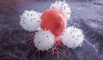
T cell killing assays quantify the ability of cytotoxic T lymphocytes (CTLs) to recognize and eliminate tumor cells. This ability represents a key effector mechanism underlying many T cell–based immunotherapies, including checkpoint inhibitors, CAR-T cells, bispecific T cell engagers, and cancer vaccines.
These co-culture systems model essential interactions between effector and target cells and allow for the quantitative assessment of the magnitude, kinetics, and antigen specificity of T cell–mediated cytotoxicity. Although they only partially replicate the complexity of the tumor microenvironment, they provide a controllable, scalable platform for evaluating how therapeutic candidates modulate T cell function. This supports prioritizing candidates before conducting more complex in vivo studies.
Evaluate anti-PD-1, anti-PD-L1, anti-CTLA-4, and next-generation checkpoint antibodies for enhanced T cell–mediated killing
Assess BiTE molecules and multispecific antibodies that bridge T cells to tumor targets via CD3 engagement
Quantify on-target killing efficacy of engineered T cells across different constructs and manufacturing conditions
Identify synergistic combinations using Bliss factor analysis for checkpoint inhibitors with targeted agents or chemotherapy
xCELLigence impedance monitoring delivers continuous data with readouts every minute—revealing killing dynamics that endpoint assays miss. Track T cell-mediated cytotoxicity over hours to days for complete kinetic profiles.
Combine cytotoxicity data with immunophenotyping (standard or custom-tailored flow panels), cytokine profiling (MSD multiplex), and T cell activation/exhaustion markers in unified study designs.
Progress from high-throughput 2D screening to physiologically relevant 3D spheroid models within a single platform.
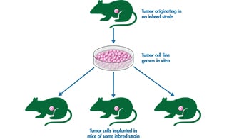
Seamlessly advance promising candidates into syngeneic or humanized mouse models with integrated immunophenotyping endpoints.
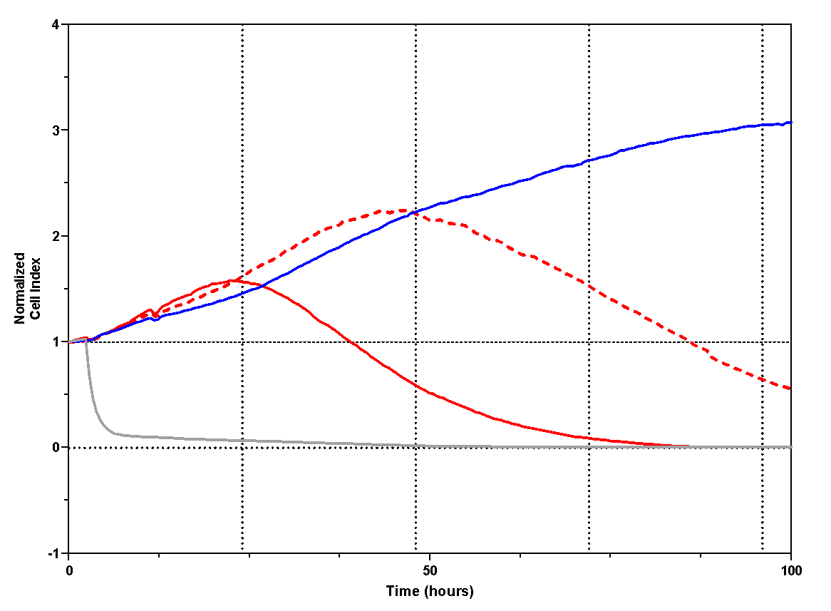
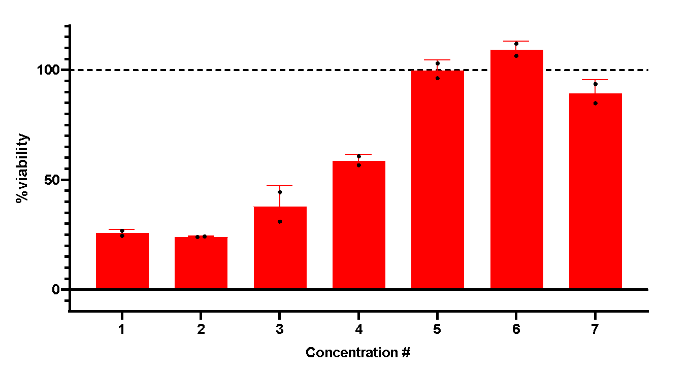
T cells are isolated from healthy human PBMCs and co‑cultured with SAOS‑2 target cells. Activation is induced using stimuli such as ImmunoCult™. Target‑cell viability is continuously monitored by impedance‑based measurement using xCELLigence technology.
ImmunoCult™ treatment (red, solid and dotted lines) induces concentration‑dependent target‑cell killing over time in co‑culture (A). The concentration‑dependent effect of ImmunoCult™ on target‑cell killing is summarized in panel B.
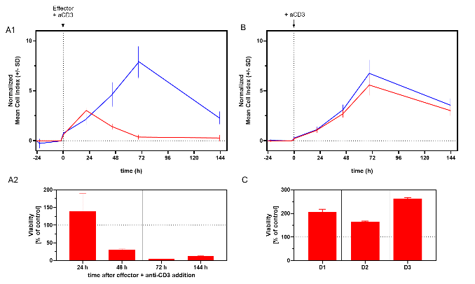
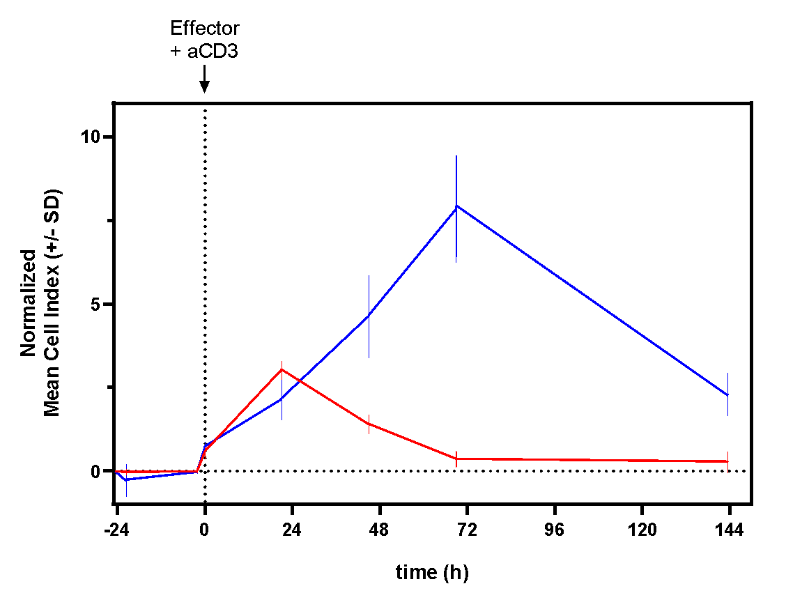
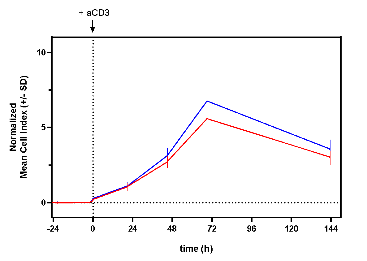
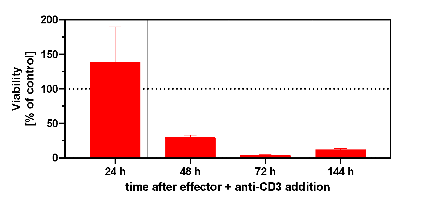
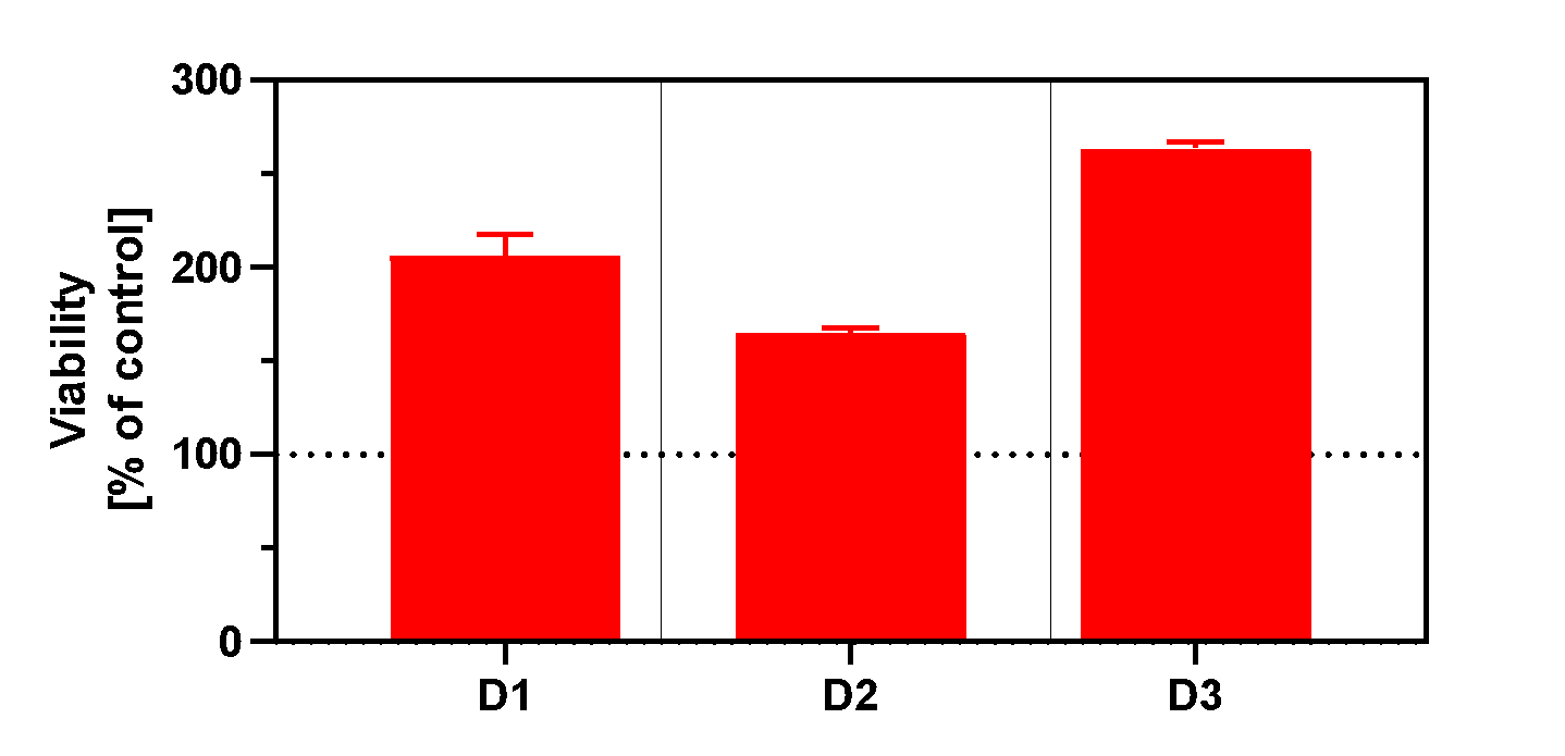
PBMCs are co-cultured with target cells and stimulated by anti-CD3 treatment (positive control). Target cell viability is assessed via impedance measurement (xCELLigence technology) over time.
Anti‑CD3 treatment (red) enhanced target‑cell killing over time in co‑culture (A1, A2). In monoculture, target cells remained unaffected (B), while effector‑cell numbers increased after 72 hours of stimulation (C, three donors).
How it could look like
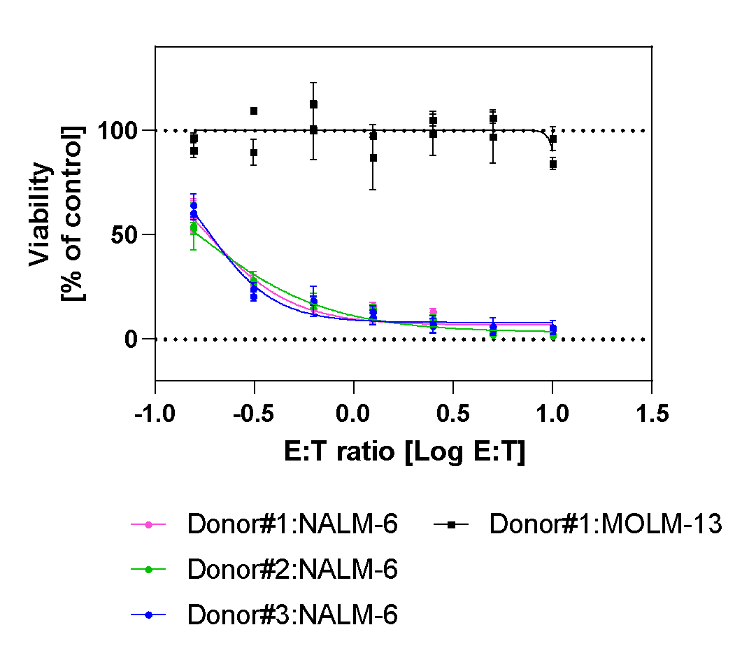
Different anti-CD19 CAR-T cell donors are co-cultured with luciferase tagged target cells, either CD19 expressing NALM-6 or non CD19 expressing MOLM-13 cells (negative control) at different effector to target cell ratios (E:T ratio). Target cell viability is assessed via luciferase activity after 72h of incubation.
Robust and selective efficacy: CD19 CAR T cells eliminated CD19⁺ NALM‑6_luc targets in all donors, while sparing CD19⁻ MOLM‑13_luc control cells.
TNF is involved in the pathology of several inflammatory and autoimmune diseases, and the use of neutralizing antibodies or engineered TNFR fusion proteins to block TNF/TNFR binding has proven to be a viable strategy for providing clinical benefits in inflammatory diseases. TNF-producing cells could be targeted for destruction, which could help reduce the inflammatory burden even more. TNF-targeted antibodies can activate effector functions such as antibody-dependent cell-mediated cytotoxicity (ADCC) or complement-dependent cytotoxicity (CDC) to destroy mTNF-expressing inflammatory cells.
Complement-dependent cytotoxicity (CDC) assays are conducted to evaluate new therapeutics during the drug development phase. The CDC assay is used for safety profiling by screening therapeutic candidates to ensure that the compound does not induce complement events and to look for other unexpected effects. Fresh blood that is less than four hours old is used in this assay, as it generates better responses and results.
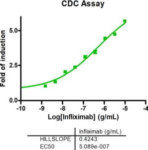
CDC activity of infliximab assessed using the CDC assay developed by Reaction Biology.
Engaging the T-cell receptor complex leads to activation of resting T cells and induction of cytokine release. In this study example, we determined the potency of drugs enhancing T cell activation by co-cultivating PBMCs with tumor cells in the presence of anti-CD3. Further, we modulated the T cell’s killing capacity with the immune checkpoint inhibitor anti-PD1.

Tumor cells stably expressing luciferase were co-cultured with PBMCs at an E:T (effector to target cell ratio) of 6:1 or a range of E:T ratios, in the presence of anti-CD3 at low concentrations to induce killing at a suboptimal level. In a second experiment, the checkpoint inhibitor anti-PD1 was added at different concentrations (7x semi-log). The co-culture was incubated for 4 days after which the viability of the tumor cells was measured by luciferase activity.
T cell stimulation via anti-CD3 antibody resulted in activation of T cells and killing of tumor cells (A). This effect was enhanced by drug combination testing with anti-PD1 antibodies (B).
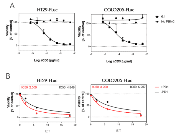
PBMCs from a healthy donor were co-cultured with cancer cell lines HT29 and COLO205 stably transduced with Firefly luciferase (FLuc).
A. An E:T ratio of 6:1 was used with the addition of semi-log dilutions of anti-human CD3 (OKT3).
B. Anti-human CD3 (OKT3) at a concentration of 0,68 ng/ml was used to stimulate T cells in a killing assay with different E:T ratios. Anti-PD1 antibody Nivolumab treatment slightly enhanced tumor cell killing.
The % viability was calculated using tumor cells alone as maximal viability (100%) and Staurosporine treatment as maximal killing (0%).
Bi-specific antibodies contain two binding sites to engage T cells and tumor cells resulting in tumor cell killing by (i) bringing the cells in close proximity, and (ii) stimulating cytokine release in T cells.
In this study example, we determine the capacity of a bi-specific engaging molecule to facilitate tumor cell killing by T cells and the combinatorial potential with other clinically relevant agents.

CD19-expressing B cell lymphoma cells stably expressing firefly luciferase are co-cultured with PBMCs at an E:T (effector to target cell ratio) of 2:1. We tested the bi-specific T cell engager Blinatumomab recognizing CD19 and CD3 at different concentrations (7x semi-log), with or without additional compounds. The co-culture was incubated for 4 days, after which the viability of the tumor cells was measured by luciferase activity.
BiTE Blinatumomab and the small molecule Lenalidomide act synergistically to kill CD19-positive B cell lymphoma cells, whereas the combination with S63845 acts antagonistically.
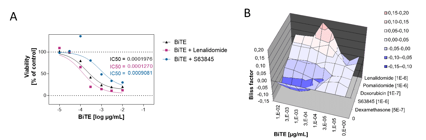
A. PBMCs from a healthy donor were incubated with the B cell lymphoma cell line OCI-LY1 at a 2:1 E:T ratio and treated with a range of 7 concentrations of Blinatumomab. Luciferase expression of live tumor cells was measured after 4 days, and viability was calculated using Staurosporine treated cells as maximal cell death control and untreated target cells as maximal viability control. An IC50 of 0,19 ng/ml was found for Blinatumomab. The combinatorial effects of Blinatumomab with Lenalidomide and MCL1-inhibitor S63845 are shown as examples.
B. Using the bliss factor scoring, we determined which drug combinations produce additive, synergistic, or antagonistic effects for killing tumor cells.
We offer assays using total PBMCs, purified CD8+ T cells, CD4+ T cells, or customer-supplied engineered cells (CAR-T, TCR-T). Cells are sourced from healthy donors with options for donor selection based on HLA type or other criteria.
We maintain an extensive library of characterized human and murine tumor cell lines across indications including solid tumors (lung, breast, colon, melanoma) and hematological malignancies. Luciferase-expressing variants and cells engineered with specific tumor-associated antigens are available. Custom lines can be generated upon request.
We recommend testing across multiple donors (minimum 3) to capture biological variability. Statistical analysis includes donor as a variable, and we can identify consistent responder profiles for follow-up studies.
Yes, we routinely assess combination treatments and calculate synergy using Bliss independence methodology. This is particularly valuable for checkpoint inhibitor combinations with targeted therapies or chemotherapy.
Standard studies are completed within 4–6 weeks from project initiation. Expedited timelines are available for priority programs.
In vitro killing assays provide rapid, mechanistic insights that help prioritize candidates before in vivo testing. We recommend using in vitro data to select top candidates for subsequent syngeneic or humanized mouse model studies within our integrated platform.
Our scientists will design a customized T cell killing assay strategy aligned with your program goals.