Soft Agar Colony Formation Assay
The standard format for adherent solid tumor cell lines. Cells grow in a 0.4% top agar layer over a 0.6% base layer in 96-well plates, with colony growth quantified by fluorescence. Performed in Freiburg, Germany.

Quantify anchorage-independent growth in solid tumor and leukemia cell lines.
Our colony formation assay service measures how your compound blocks single tumor cells from growing into colonies. Run adherent solid tumor lines in soft agar, or suspension and leukemia lines in methylcellulose, with IC50 and combination readouts.
Normal epithelial cells need attachment to a basement membrane to survive; detached, they undergo anoikis. Transformed cells escape this control and keep dividing in suspension. A colony formation assay, also called a clonogenic assay, measures this anchorage-independent growth by suspending single cells in a semi-solid matrix, where only clonogenic cells expand into colonies.
Because the readout tracks a hallmark of transformation rather than bulk proliferation, the assay can reveal compound activity that 2D viability assays miss. We run two formats matched to cell type: soft agar for adherent solid tumor lines and methylcellulose for suspension and leukemia lines.
The standard format for adherent solid tumor cell lines. Cells grow in a 0.4% top agar layer over a 0.6% base layer in 96-well plates, with colony growth quantified by fluorescence. Performed in Freiburg, Germany.
The preferred format for suspension and leukemia cell lines. A 1% methylcellulose matrix immobilizes single cells for 14 days of clonal expansion, with automated colony counting and sizing. Performed in Malvern, Pennsylvania.
Both formats support combination treatment alongside single-agent IC50 determination, so combination effects are measured on clonogenic outgrowth, not only on 2D viability.
Working with an adherent line not yet established in our soft agar format? We can set up the assay with your cell line on request.
| Parameter | Soft Agar | Methylcellulose |
| Best for | Adherent solid tumor cell lines | Suspension and leukemia cell lines |
| Matrix | 0.4% agar over a 0.6% agar base layer | 1% methylcellulose in complete growth medium |
| Plate format | 96-well | 96-well |
| Culture period | Until colonies form in the solvent control; up to 3 weeks of compound exposure | 14 days at 37°C |
| Readout | Resazurin viability fluorescence | MTT stain with Oxford Optronix GelCount automated imaging |
| Output | IC50; percent colony growth vs. controls | IC50; colony number and size distribution |
| Screening facility | Freiburg, Germany | Malvern, Pennsylvania |
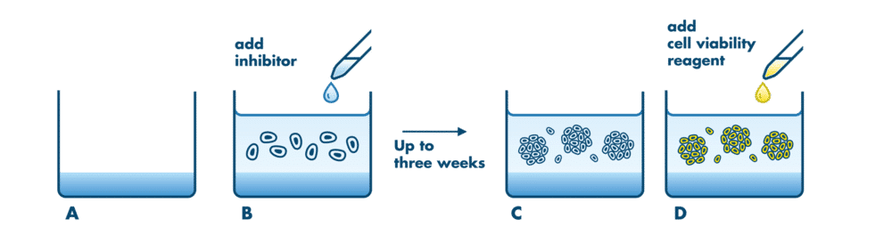
Single cells are seeded in soft agar medium for growth independent of the attachment to a surface. Tumor cells, as opposed to other non-transformed cell types, can divide in an anchorage-independent way with single cells building cell colonies over a course of several days or a few weeks.
Soft agar for adherent solid tumor lines and methylcellulose for suspension and leukemia lines, so blood and solid tumor programs get the format each cell type actually grows in.
Up to three weeks of compound exposure in soft agar and 14 days in methylcellulose, giving compounds time to act on clonal outgrowth that a 3-day 2D assay can miss.
Every study includes IC50 determination of a reference compound alongside your test articles, so each data set carries its own benchmark for assay performance.
Our soft agar format was established on 60 tumor cell lines with Z’ factors above 0.4, supporting compound profiling across many cell lines in one study.
Methylcellulose plates are scanned on the Oxford Optronix GelCount imaging system, which reports both colony number and colony size distribution for each condition.
Single cells are suspended in a semi-solid matrix that prevents attachment while letting nutrients and test compounds diffuse freely.
Test compounds are added across a concentration range, alone or in combination.
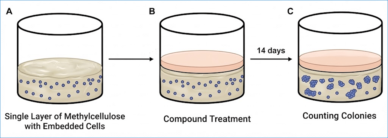
Cells incubate until colonies form in the solvent control, typically 1 to 3 weeks depending on cell line and format.
Colony growth is quantified by viability staining and fluorescence (soft agar) or automated imaging of colony number and size (methylcellulose).
Results are reported as percent colony growth relative to controls, with IC50 values from sigmoidal dose-response fitting.
Approach
The Aurora B inhibitor VX-680 was tested for inhibition of soft agar growth of A549 non-small cell lung cancer cells.
Setup
Wells of a 96-well plate are coated with 0.6% soft agar, followed by seeding of the cells in 0.4% soft agar. After the agar solidifies, compounds are added and cells incubate until colonies form in the solvent control. A resazurin viability reagent is then added, and fluorescence is measured as an indirect quantification of colony growth.
Results
Colonies formed for 5 days, were photographed and stained with a fluorescent viability agent. Fluorescence was expressed as percent colony growth relative to solvent alone, giving an IC50 of 1.6×10⁻⁷ M.

Approach
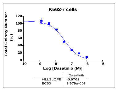
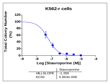
Drug-resistant K562 cells (K562-r) were treated with dasatinib or staurosporine to measure inhibition of clonogenic growth in methylcellulose.
Setup
Suspension cells are mixed with 1% methylcellulose in complete growth medium and seeded into 96-well plates. Compounds are added and cultures incubate at 37°C for 14 days to allow colony formation. MTT is added to visualize viable colonies, and plates are scanned on the Oxford Optronix GelCount imaging system for automated quantification of colony number and size distribution.
Results
IC50 values were calculated in GraphPad Prism by fitting colony counts to a sigmoidal dose-response model. Dasatinib reduced colony number with an IC50 of 3.98×10⁻⁸ M; staurosporine with an IC50 of 5.96×10⁻⁸ M.
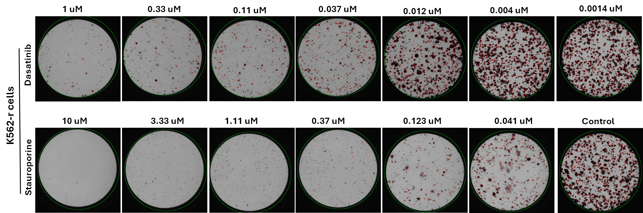
Representative images of K562-r colonies after 14 days of treatment with dasatinib or staurosporine.
Also called a clonogenic assay, it measures the ability of single cells to grow into colonies. In semi-solid formats like soft agar and methylcellulose, cells cannot attach to a surface, so colony growth reflects anchorage-independent growth, a hallmark of malignant transformation. Reduced colony formation after treatment is a sensitive endpoint for anticancer activity.
Soft agar is the standard format for adherent solid tumor cell lines. Methylcellulose is preferred for suspension cells, including leukemia cell lines, because its viscous matrix immobilizes single cells and supports their clonal expansion. If your program spans both tumor types, we can run each cell line in its appropriate format.
A colony formation assay tests whether a compound stops single cells from growing into colonies. A 3D tumor spheroid assay tests a compound’s anti-proliferative and cytotoxic effect on spheroids that have already formed. Colony formation addresses clonogenic outgrowth; spheroid assays address established multicellular tumor structures.
In our soft agar profiling across 60 tumor cell lines, some compounds were markedly more potent in soft agar than in 2D proliferation assays. Transformed cells can be more sensitive to transformation-specific inhibitors when growing anchorage-independently, so soft agar can pick up activity that a 2D assay misses.
Yes. Both soft agar and methylcellulose formats support drug combination testing in addition to single-agent IC50 determination.
For soft agar, 30 µl of a stock solution at 1000x the highest assay concentration, or the equivalent solid material. For methylcellulose, 50 µl of a 1000x stock or 2 mg of solid material. Our FAQ page has the cell-based assay condition form with preparation and shipping details.
Methylcellulose studies have a 4 to 5 week turnaround, and expedited scheduling can be arranged before the study starts. Contact us for soft agar timelines for your cell lines.